CORONAVIRIDAE - PowerPoint PPT Presentation
1 / 56
Title:
CORONAVIRIDAE
Description:
III (avian) Infectious bronchitis virus of Tracheobronchitis, nephritis ... Outbreaks of infectious bronchitis have declined in recent years through use of ... – PowerPoint PPT presentation
Number of Views:2958
Avg rating:3.0/5.0
Title: CORONAVIRIDAE
1
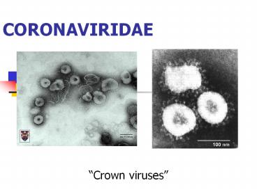
CORONAVIRIDAE
- Crown viruses
2
General Characteristics- Coronaviruses
- Single-stranded, positive sense RNA viruses
- Enveloped and pleomorphic
- Infect a wide range of mammalian and avian
species - Cause respiratory and enteric disease,
encephalomyelitis, hepatitis, serositis, and
vasculitis in domestic animals. In humans, common
cold. - Pronounced tropism for epithelial cells of the
respiratory and the intestinal tract. - In general, mild or inapparent infections in
adults but severe diseases in newborn or young
animals. - Stability of virus cold temperature relatively
stable labile at room temperature highly
photosensitive
3
Viruses with ve RNA genomes
foot and mouth disease virus
Picornaviridae
porcine enteroviruses
Caliciviridae
Coronaviridae
feline calicivirus
coronaviruses
Arteriviridae
equine arterivirus, PRRSV
Flaviviridae
flaviviruses (WNV)
pestiviruses (BVD)
Togaviridae
equine encephalitis viruses
4
Classification and General Characteristics
- Coronaviruses have also been associated with
infections of the respiratory and enteric tracts
and with central nervous system disease in
monkeys, rats, rabbits, and other species. - Unusually large club-shaped peplomers projecting
from the envelope give the particle the
appearance of a solar corona pleomorphic 75-160
nm - Family Coronaviruses have been divided into four
antigenic groups (Table 1). Viruses within each
group show some antigenic cross-reactivity, and
there may be a number of serotypes within one
virus species. Animals immune to one serotype are
susceptible to infection with different serotypes
of the same coronavirus.
5
Table 1. Antigenic Groups and Diseases Caused by
Coronaviruses
Antigenic Group Virus Disease
I (mammalian) Human coronavirus 299E Common
cold Transmissible gastroenteritis Gastroenteriti
s Feline infectious peritonitis Peritonitis,
pneumonia virus, meningoencephalitis, pano
phthalmitis, wasting Canine coronavirus Enteritis
II (mammalian) Human coronavirus OC43 Common
cold Mouse hepatitis virus (many Hepatitis,
encephalomyelitis, serotypes) enteritis Bovine
coronavirus Gastroenteritis Porcine
hemagglutinating Vomiting, wasting,
and encephalomyelitis virus encephalomyelitis
III (avian) Infectious bronchitis virus
of Tracheobronchitis, nephritis chickens (3
eight serotypes) IV (avian) Bluecomb disease
virus of Enteritis turkeys
6
Coronavirus Structure
- The envelope carries three glycoproteins
- S - Spike protein receptor binding, cell fusion,
major antigen - E - Envelope protein small, envelope-associated
protein - M - Membrane protein transmembrane - budding
envelope formation In a few types, there is a
third glycoprotein - HE - Haemagglutinin-esterase
- The genome is associated with a basic
phosphoprotein, N.
7
Coronavirus replication. Numbers of mRNAs and
locations of nonstructural (NS) proteins may vary
for different cornaviruses. Virions bind to the
cell membrane and enter by membrane fusion or
endocytosis. Viral genomic RNA acts as mRNA to
direct the synthesis of viral RNA-dependent RNA
polymerase. This enzyme copies the viral genomic
RNA to form full-length (-) strand templates.
These templates are copied to form new () strand
genomic RNA, an overlapping series of subgenomic
mRNAs, and leader RNA. All mRNAs are capped and
polyadenylated and form a nested set with common
3 ends. Each mRNA codes for a single
polypeptide. The N protein binds to novel viral
RNA to form helical nucleocapsids. E1, E2, and
E3 glycoproteins are produced on membrane-bound
polysomes. Some coronaviruses do not encode E3.
Cornaviruses that encode E3 cause hemadsorption
in infected cells. Virions are formed by budding
at membranes of the Golgi apparatus and the RER,
but not at the plasma membrane. Virions are
released by cell lysis or by fusion of
post-Golgi, virion-containing vesicles with the
plasma membrane.
8
Replication
9
SARS Severe Acute Respiratory Sydrome
- February 2003 Guangdong, China
- Viral pneumonia, fever cough,
- dyspnea, headache, and hypoxemia
- High case fatality
Lipsitch et al 2003, Science 300 1966
10
SARS Coronavirus
11
SARS
Where did SARS come from?
palm civet (paguma larvata)
12
SARS diagnosis
Serology ELISA, questionable Virology-PCR
13
Viruses of Veterinary Importance Coronaviruses
- Bovine coronavirus (BCV)
- Transmissible gastroenteritis (TGE)
- Feline infectious peritonitis (FIP)
- Infectious bronchitis disease virus (IBDV)
14
Bovine Coronavirus (BCV)
- Viral diarrhea- Rotaviruses are the major cause
of diarrhea in the young calf. Coronaviruses are
also important. The pathogenesis is similar
between the two viruses - Disease is most commonly seen in calves at about
1 week of age, the time when antibody in the
dam's milk has fallen to a low level. The
diarrhea usually lasts for 4 or 5 days. The
destruction of the absorptive cells of the
intestinal epithelium of the small intestine, and
to a lesser extent those of the large intestine,
leads to the rapid loss of water and
electrolytes. - Glucose and lactate metabolism is affected
hypoglycemia, lactic acidosis, and hypervolemia
can lead to acute shock, heart failure, and
death, although coronavirus diarrhea is generally
less severe than that caused by rotaviruses.
Bovine coronaviruses may cause diarrhea in humans.
15
Viral causes of diarrhea in neonates
- Rotavirus
- Coronavirus
- BVD
- Bredavirus
- Calicivirus
- Parvovirus
- Astrovirus
16
Susceptability of neonates
- Rotaviruses 4 to 14 days
- Coronavirus 4 to 30
4 days
0
Colostral Antibodies in gut
Susceptible period
17
Diagnosis
- FA of fecal samples
- EM
18
Prevention
- Vaccination of pregnant animals
- Colostrum for 2 weeks
19
vaccines against calf diarrhoea
20
Bovine Coronavirus (BCV)
- Winter dysentery is a sporadic acute disease of
adult cattle that occurs in many countries
throughout the world, and it is believed to be
caused by coronaviruses. - The clinical syndrome is characterized by bloody
diarrhea accompanied by decreased milk
production, depression, and anorexia. - Available vaccines are not effective, because
they do not appear to contain sufficient
antigenic mass and cannot be given early enough.
Alternatives to vaccinating calves are to
immunize the dam to promote elevated antibody
levels in the colostrum or to feed antibody
directly to the calf in colostrum.
21
Transmissible Gastroenteritis Virus (TGEV)
22
Transmissible Gastroenteritis Virus (TGEV)
- TGEV of swine usually occurs in the winter months
- Characterized by an explosive outbreak of
vomiting and profuse diarrhea - Transmissible gastroenteritis is one of the major
causes of death in young piglets in the a
midwestern United States. Mortality is high,
vaccines are of limited efficacy, and it appears
to be difficult to prevent the introduction of
the virus into herds.
23
Transmissible Gastroenteritis Virus (TGEV)
- Clinical
Features - The disease is usually recognized at farrowing
time. - The incubation period is usually 1-3 days, and
all litters within the farrowing house are
commonly affected at the same time. - The clinical signs in piglets are vomiting
followed by a watery diarrhea and rapid loss of
weight. The diarrhea is profuse, with an
offensive odor, and often contains curds of
undigested milk. - Piglets infected when under 7 days of age
generally die within 2 to 7 days of the onset of
signs piglets over 3 weeks of age usually live
(may be unthrifty for several weeks). In growing,
finishing, and adult swine the disease is
commonly associated with inappetence and diarrhea
of a few days' duration, and may even go
unnoticed. Sows infected late in pregnancy may
develop pyrexia, but they are otherwise normal
and rarely abort.
24
(No Transcript)
25
(No Transcript)
26
(No Transcript)
27
TGE IHC Immunocytochemistry Infected
Epithethial Cells
28
Transmissible Gastroenteritis Virus (TGEV)
- Diagnosis
- presumptive diagnosis of TGE can be made from
the sudden appearance of a rapidly spreading and
often fatal disease of young piglets accompanied
by vomiting and diarrhea. - clinical diagnosis can be confirmed by
demonstration of specific antigen by
immunofluorescence, isolation of virus, and
demonstration of rising antibody titers in paired
sera.
29
Transmissible Gastroenteritis Virus (TGEV)
- Epidemiology and Control
- Transmissible gastroenteritis occurs most
commonly in the winter months (in North America
between November and April), but its source is
unknown. Its presence becomes apparent only when
large numbers of piglets are born at a time when
weather conditions favor transmission. - Control is difficult, although good management of
the farrowing house can reduce the risk. The most
widely used vaccination regimen involves
vaccinating the sow with an attenuated vaccine 3
weeks before farrowing, thus providing piglets
with high levels of protective antibody in the
colostrum during the critical first few days of
life.
30
feline infectious peritonitis
Horzinek and H. Lutz An update on FIP Veterinary
Sience Tomorrow Jan, 2001 www.vetscite.org
31
FIP
- fatal disease of young cats (3-18 mo) in
multi-cat houses or catteries - not seen before 1950
- new virus?
- old virus, new disease
- systemic antibodies not protective, may even be
harmful (antibody dependent enhancement, early
death)
32
feline enteric coronavirus
- closely related to dog, pig (TGE), human
coronaviruses - species specific but K9CV can infect cats
- two serotypes
- serotype I
- more common, 70-95 of isolates, does not cross
react with K9CV - difficult to isolate
33
FeCV, serotype 2
Both serotypes can lead to FIP causing strains
34
FeCV
- very prone to making mistakes during replication
- 1/10,000 nucleotides
- quasispecies
- invariant portion of genome
- primers for RT-PCR
- mild enteric or respiratory disease
- grows mainly in epithelial cells
- persistent infections
- in balance with immune system
- low levels of antibody
35
FIPvirus
- derived by mutation from FeCV
- nature of mutation not defined
- few obvious common mutations in FIP causing
strains - No reliable technique for differentiating between
non-virulent and virulent strains - not usually spread from cat to cat
36
epidemiology
- Exposure to FeCV
- 25 of cats from 1-2 cat households are
seropositive - 75-100 of cats from catteries seropositive
- susceptible cats become infected immediately
following exposure - kittens can become infected in utero or soon
after maternal antibodies drop below protective
levels
37
epidemiology (FIP)
- 15,000 in 1-2 cat households
- 120 in catteries
- sporadic
- clustered9 (2-3 cats)
- rarely epidemic - 40 mortality
- no gender or breed predisposition
38
persistent infections
- cats can shed virus (RT-PCR of blood and feces)
for long time - persistence not reinfection
- virus replicates in a few epithelial and lymphoid
cells - immunohistochemistry
- each cat has own collection of viruses
- protected from infection by other strains
- premunition
- reason for rare horizontal transmission of FIP
39
FIP pathogenesis
FEC
Mild diarrhoea or respiratory illness
virus
immune system
persistent infection
low level of replication in epithelial and
lymphoid cells
40
stress
pregnancy in young queens
elective surgery
concurrent infections (FeLV, FIV ?)
weaning, sale, shipment, adaptation
41
Virus
immune system
increased virus replication -gt virulent mutants
increased ability to grow in macrophages
immune-mediated lysis of infected cells
cytokines draw in more susceptible cells
vascular permeability
immune complex related damage
42
(No Transcript)
43
clinical signs
- common signs
- chronic antibiotic unresponsive fever
- progressive anorexia, weight loss
- stunting of growth
- progressive increase in serum proteins
- increase in globulins
- anemia
- serum, urine brown due to bilirubin
44
clinical signs
- wet form
- peritonitis
- pleuritis
- dry form
- surface oriented granulomas
- mesenteric lymph nodes, liver, kidneys, cecum
(palpable) - cloudiness in eye
- neurological signs
- can change from dry to wet
45
diagnosis
- serology
- prognosis?
- no titer - no FIP but may still be infected
- lt100 - less chance of developing FIP
- gt100 - greater chance of getting FIP
- increased globulins and protein (gt35g/L)
- cytology
- degenerate and non-degenerate PMN, macrophages,
some lymphocytes, protein background - FeCV positive cells (FAT)
46
diagnostic alogrithm (Horzinek and Lutz)
47
diagnostic algorithm
48
diagnostic algorithm
49
control
- vaccine
- Primucell FIP
- Intranasal ts virus
- management
- early weaning and separation
50
Viruses of Veterinary ImportanceAvian Infectious
Bronchitis Virus (IBV)
- Avian infectious bronchitis (gasping disease) is
one of the most important viral diseases of
chickens. IBV is responsible for an acute
respiratory disease which can produce very high
mortality rates in young chicks.
51
Viruses of Veterinary Importance Avian
Infectious Bronchitis Virus (IBV)
- Clinical Features
- Outbreaks of infectious bronchitis are explosive.
IBV spreads rapidly to involve the entire flock
within a few days. - Chicks between 1 and 4 weeks of age show the most
severe disease, which is recognized initially by
coughing, sneezing, nasal discharge, and
respiratory distress. Mortality in young chicks
is usually 25-30 but in some outbreaks can be as
high as 75. - In older birds the disease often goes unnoticed,
but in laying hens there is a marked drop in egg
production, with many soft-shelled and malformed
eggs being laid.
52
Viruses of Veterinary Importance Avian
Infectious Bronchitis Virus (IBV)
- Pathology and Pathogenesis
- The course of the disease in young chicks is from
7 to 21 days depending on the severity of the
disease. Necropsy of young chicks dying from
infectious bronchitis shows sinuosities,
catarrhal tracheotis, bronchitis, and congestion
and edema of the lungs. Caseous plugs may be
present in the bronchi. - The primary target for viral replication is the
trachea, but the virus also replicates in the
lungs, ovaries, and lymphoid tissue. - IBV can establish persistent infection in some
chickens, which results in shedding of virus in
the feces for several months after initial
exposure to the virus. When virus persists in the
presence of high levels of antibody, severe
nephritis can occur, which possibly reflects an
immune complex-mediated disease.
53
Viruses of Veterinary Importance Avian
Infectious Bronchitis Virus (IBV)
- Laboratory Diagnosis
- In contrast to several of the coronaviruses, IBV
can be easily isolated by the allantoic
inoculation of 9- to 12-day-old embryonated eggs
obtained from seronegative hens. Infected embryos
are to a variable degree stunted or curled
tightly. A range of cell and organ cultures can
also be used for virus isolation. - At least eight genotypes of IBV exist and fall
into two major groups virus isolates of widely
differing pathogenicity occur within each
antigenic group.
54
Avian Infectious Bronchitis Virus (IBV)
- Epidemiology and Control
- IBV spreads between birds by aerosol and by
ingestion of food contaminated with feces.
Control of infectious bronchitis is difficult
because of the presence of persistently infected
chickens in many flocks. - Outbreaks of infectious bronchitis have declined
in recent years through use of vaccines however,
it may occur even in vaccinated flocks following
the introduction of infected replacement chicks
from another farm. To minimize this risk, most
poultry farms purchase only 1-day-old chicks and
rear them in isolation. - Attenuated vaccines, administered in the drinking
water or as aerosols, are widely used to protect
chicks and are usually given between 7 and 10
days, and again at 4 weeks. Vaccination earlier
than 7 days may be unsuccessful because most
chicks have passive immunity up to this age. - Local immunity in the respiratory system is
critical for protection and can be generated by
heterotypic vaccine strains.
55
Avian infectious bronchitis. (A) One synonym for
the disease is gasping disease. (B) Thick
mucopurulent exudate in the trachea. (C)
Nephrosis. The kidney is pale and enlarged to
about five times normal size. (D) Embryos from
embryonated hens eggs inoculated via the
allantoic cavity with serial dilutions of virus
when 9 days old, and examined 11 days later.
Amounts of virus diminish in pairs from right to
left in the top row, and from left to right in
the bottom row.
56
Other coronaviruses of importance in veterinary
medicine
- Porcine respiratory coronavirus (PRCV)
- Porcine Hemagglutinating Encephalomyelitis virus
- Porcine epidemic diarrhea virus (PEDV)
- Canine coronavirus (CCV)
- Turkey coronavirus (Bluecomb disease of turkeys)
- Mouse Hepatitis Virus


