Presentazione di PowerPoint - PowerPoint PPT Presentation
1 / 1
Title:
Presentazione di PowerPoint
Description:
... study of nanocrystallization in amorphous matrix for non-linear ... The as-quenched sample is amorphous and the surface appears almost flat (Fig.1 a) ... – PowerPoint PPT presentation
Number of Views:18
Avg rating:3.0/5.0
Title: Presentazione di PowerPoint
1
Structural and morphological study of
nanocrystallization in amorphous matrix for
non-linear optical applications
Paolo Colombia, Paolo Bergesea, Elza Bontempia,
Pasquale Perniceb, Antonio Aronneb, Vladimir N.
Sigaevc, Laura E. Deperoa.
a) INSTM and Structural Chemistry Laboratory,
University of Brescia, Via Branze, 38, 25123
Brescia, Italy b) Department of Materials and
Production Engineering, University of Naples
Federico II, P. le Tecchio, 80125 Naples,
Italy c) Mendeleev University of Chemical
Technology, Miusskaya Square,9, 125190 Moscow,
Russia colombi_at_ing.unibs.it
Second harmonic generation (SHG) in glass and
glass ceramics opens new frontiers in optical
material science. In spite of the fact that
macroscopic inversion symmetries theoretically
exclude SHG in glasses (e.g. isotropic media),
observations of SHG in glassy materials have been
reported. A possible explanation of this
behaviour is that, in such materials, non-linear
optical activity is connected to the nucleation
in the glassy matrix of non-centrosymmetric
ferroelectric nanocrystallites. Therefore, many
attempts have been made in order to grow
nanocrystallites of ferroelectric or other highly
polarizable phases in glassy materials. The
synthesis and structural characterisation of
nanostructured potassium niobium silicate (KNS)
glasses xK2O xNb2O5(1-2x)SiO2 have been recently
reported V.N. Sigaev et al.Non-Cryst. Solids 306
(2002) 238 In the present work, nanostructuring
in the amorphous matrix of KNS glasses is
investigated by means of atomic force microscopy
(AFM) and 2D X-Ray diffraction (XRD2). Synergic
application of AFM imaging and XRD analysis
allowed us to correlate morphological and
structural information and in turn to shed some
light on the influence of the thermal treatment
on the growth of the nanocrystals in the matrix.
a)
d)
c)
b)
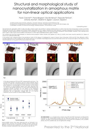
Fig 1. In the above sequence AFM and XRD2
measurements are shown. The as-quenched sample
is amorphous and the surface appears almost flat
(Fig.1 a). After 24 h of thermal treatment at 953
K a nanocrystlline phase appears in
correspondence to that treatment on the surface
the presence of nanocrystallites clusters have
been detected (Fig. 1c). Since in the sample
treated for 70 h at the same temperature the
integrated intensity increases, but the FWHM of
the integrated diffraction peak is similar (Fig.
2), we conclude that the crystallite dimensions
do not significantly change after 24 h of thermal
treatment, but the number of nanocrystallites
increases.
IN CONCLUSION, by a complementary analysis using
AFM and XRD2 techniques we followed the formation
of the nanocrystlline phases and we observed
clusters both on cut and fracture surfaces. The
effect of the thermal treatment time is to
promote the nucleation of the crystalline phase,
since the crystalline sizes do not significantly
change.
Fig. 3
Nanocrystallite clusters have been detected both
on fractured a cut surfaces (Fig.3). However,
the average roughness of these surfaces are
different. In particular the roughness of cut
surfaces are always larger. This effect has been
attributed to the mechanical cut (see Fig. 1b)
Presented to the 2nd National Conference on
Nanoscience and Nanotechnology The Molecular
Approach Area del CNR di Bologna 25/27
Febbraio 2004


