BIO 148 Lab Test 2 - PowerPoint PPT Presentation
1 / 80
Title:
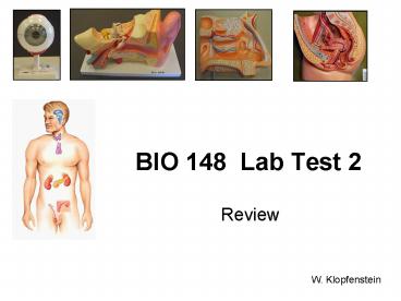
BIO 148 Lab Test 2
Description:
Do not use these PowerPoint images in lieu of going to lab! ... (structure with striated appearance) (hole) (layer seen wrinkled here) (circular area) ... – PowerPoint PPT presentation
Number of Views:75
Avg rating:3.0/5.0
Title: BIO 148 Lab Test 2
1
BIO 148 Lab Test 2
- Review
W. Klopfenstein
2
Caution!
- Do not use these PowerPoint images in lieu of
going to lab! If you do not actually view the
slides, models, dissections, and other lab
materials first-hand in the lab your chances of
doing well on the lab test are likely to be poor.
- The lab test will not be constructed using images
of this PowerPoint presentation. - This presentation does not necessarily review
every aspect of the lab exercises on which you
will be tested. Be aware of the objectives
listed in the lab manual and any announcements
made by your lab or lecture instructor as to test
content. - This should not be you only method of reviewing
for the lab test. For example you should also
reread all of the assigned exercises in your lab
book.
3
Topics Covered
- Special Senses
- Vision
- Auditory
- Olfaction
- Endocrine System
- Reproductive System
4
Vision (the Eye)
- Eye Model
- Sheep Eye Dissection
- Retina Slide
- Eye Tests and Demonstrations
5
Eye Model
- Identify the indicated structures on the
following slides.
6
(No Transcript)
7
(hole)
8
(No Transcript)
9
(No Transcript)
10
(hole)
11
What structure fills this space behind the lens?
12
Sheep Eye Dissection
- Identify the indicated structures on the
following slides.
13
(jelly-like mass)
(hole)
(structure with striated appearance)
14
(No Transcript)
15
(layer seen wrinkled here)
(dark pigmented layer)
(circular area)
16
(No Transcript)
17
Retina Slide
- Identify the indicated structures on the
following slides.
18
(identify region)
19
Identify these there layers in the eyeball.
20
Identify the type of neurons in each of the
three indicated bands.
A. B.
Which of these two arrows shows the direction
from which light would enter the tissue layers as
they are orientated on this slide?
21
Vision Tests and Demonstrations
22
What aspect of vision does this chart test?
20/40 To what do each of
these two numbers refer when performing this
test?
20/40
23
How might this chart appear to persons with
uncorrected astigmatism? What are different
types of defects might cause astigmatism?
24
- What term describes the changing of the shape of
the lens when the eye is focused for close
vision? - Does the lens become more convex or less convex
when the eye is focused for close vision? - Does the pupil increase or decrease in diameter
when the eye is focused for close vision?
Lens
25
What reflex is being demonstrated here?
Is this reflex the result of smooth muscle or
skeletal muscle contraction?
26
What feature of vision was demonstrated in lab
using this card?
27
Auditory Sense (Ear)
- Ear Model
- Model of the Cochlea
- Auditory Ossicles
28
Ear Model
- Identify the indicated structures on the
following slides.
29
(canal)
30
(canal)
31
(No Transcript)
32
(No Transcript)
33
What sensory organ is found within this structure?
34
What membrane does this bone rest against?
35
Cross section of Cochlea
- Identify the indicated structures on the
following slides.
36
(No Transcript)
37
What is the collective term for the sensory
structure seen here?
38
Endocrine System
- Brain Model
- Torso Model
- Slide of Thyroid Gland
39
Identify each of these endocrine glands
40
The numbers below indicate the number of major
hormones secreted by each of the endocrine organs
indicated. For each gland list the names of
these specific hormones secreted and their
function.
(1)
(8)
(3)
(1)
(1)
(5)
(2)
(2)
(1)
41
Torso Models
- Identify the indicated Endocrine organs
42
This spherical structure is shown removed on this
model but is visible on the sheep brain
dissection.
43
(gland)
44
(No Transcript)
45
(No Transcript)
46
(No Transcript)
47
Complete the Following Tables of Hormones
48
(No Transcript)
49
(No Transcript)
50
(No Transcript)
51
(No Transcript)
52
(No Transcript)
53
Thyroid Slide
54
Identify this structure.
What is the name for material?
55
Identify the indicated cells and name the
hormones that they each secrete.
56
Reproductive System
- Male reproductive Models
- Slides of Male reproductive tissue
- Female reproductive Models
- Slides of Female reproductive tissue
57
Male Pelvic Model
- Identify the indicated structures on the
following slides.
58
(No Transcript)
59
Urinary Bladder
60
(No Transcript)
61
Female Pelvic Model
- Identify the indicated structures on the
following slides.
62
(No Transcript)
63
(finger-like processes)
(organ)
(region)
64
(layer)
(layer)
(cavity)
65
(No Transcript)
66
Reproductive Slides
- On the next slide identify the organs from which
these tissue samples were taken
67
3.
1.
2.
3.
4.
68
Reproductive Slides
- For each of the next slides identify the organ
from which the tissue came.
69
(cells)
From what organ was this tissue taken?
(cavity)
70
(cells)
71
(cell)
72
From what organ was this tissue taken?
73
Chart of Ovarian Follicles
- Identify which of these ovarian follicles are
- Graafian (mature)
- Primordial
- Primary
- Secondary
74
Identify the follicle type indicated by the
arrows.
From what organ was this tissue taken?
75
Identify the follicle types indicated by the
arrows.
76
Identify the follicle type indicated by the black
arrow.
(cell)
77
Identify the follicle type indicated by the black
arrow.
(cell)
78
(layer)
(layer)
From what organ was this tissue taken?
79
Identify the layers indicated by arrows.
From what organ was this tissue taken?
80


