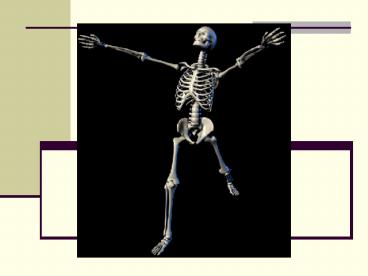
The Axial Skeleton - PowerPoint PPT Presentation
1 / 48
Title:
The Axial Skeleton
Description:
Title: PowerPoint Presentation Last modified by: student Created Date: 1/1/1601 12:00:00 AM Document presentation format: On-screen Show Other titles – PowerPoint PPT presentation
Number of Views:153
Avg rating:3.0/5.0
Title: The Axial Skeleton
1
The Axial Skeleton
- Chapter 7
2
I. Skeletal Divisions (206 bones)
- Axial Skeleton (80 bones)
- Forms longitudinal axis of the body
- Consists of
- Skull
- Vertebral column
- Ribs
- Sternum
3
- Appendicular Skeleton (126 bones)
- Consists of
- Pectoral girdle
- Pelvic girdle
- Bones of the limbs
4
II. The Skull (22 bones)
- Functions
- Protects
- Guards entrances to digestive respiratory
systems
5
- Cranium (braincase) (8 bones)
6
1. Occipital Bone (1 bone)
- Forms posterior inferior surfaces
7
2. Parietal Bones (2 bones)
- Forms superior lateral surfaces
8
3. Frontal Bone (1 bone)
- Forms anterior portion of skull roof of orbits
9
4. Temporal Bones (2 bones)
- Surrounds protects sense organs of inner ear
10
5. Sphenoid Bone (1 bone)
- Cross-brace that strengthens sides of skull
(looks like a bat)
11
6. Ethmoid Bone (1 bone)
- Forms roof of nasal cavity, part of nasal septum
12
- Facial Bones ( 14 bones)
13
1. Maxillary Bones (2 bones)
- Supports the teeth
14
2. Palatine Bones (2 bones)
- Forms portion of hard palate
15
3. Lacrimal Bones (2 bones)
- Forms medial wall of orbits
16
4. Nasal Bones (2 bones)
- Supports superior portion of bridge of nose
17
5. Zygomatic Bones (2 bones)
- Forms rim lateral wall of orbits
18
6. Vomer Bone (1 bone)
- Forms interior portion of bony nasal septum
19
7. Inferior Nasal Conchae (2 bones)
- Creates turbulence in air passing through nasal
cavity - WHY???
20
8. Mandible Bone (1 bone)
- Lower jaw
21
- Sinuses
- Makes bones lighter
- Produces mucus to moisten clean air in and near
the sinuses
22
- E. Sutures- Immovable joints connected with dense
fibrous connective tissue - Lambdoidal Suture- between occipital parietal
bones - Coronal Suture- between frontal and parietal
bones - Sagittal Suture- between parietal bones
23
- 4. Squamosal Sutures- between temporal
parietal bones - 5. Fontanels- fibrous area between cranial
bones in infants - a. allow skull to be distorted/squished to
ease delivery - b. the frontal fontanel persists until a child
is nearly 2 yrs. old
24
Sutures of the Skull
25
F. Associated Bones of the Skull (7)
- Auditory Ossicles (6 bones)
- 3 bones per ear
- malleus, incus staples
26
- Hyoid Bone (1 bone)
- Supports larynx
- Only free standing bone not connected to another
bone
27
III. The Vertebral Column
- - 33 total bones
28
A. Functions of the V.C.
- Provide a column of support
- Bear the weight of the head, neck trunk
- Protect the spinal cord
- Helps maintain an upright body position
- (Sitting/Standing)
29
B. Divisions of the V.C.
- Cervical Region
- a) Made of 7 vertebrae
- b) Constitutes the neck region
- c) Labeled C1-C7 (Superior to Inferior)
- i. C1 is called the Atlas
- -holds up the head
- - Articulates w/ occipital condyles
- - Allows yes movement
- ii. C2 is called the Axis
- - Pivots around the Atlas
- - Allows no movement
30
- Thoracic Region
- a) Made of 12 vertebrae
- b) Constitutes the chest/upper back region
- c) Labeled T1-T12 (Superior to Inferior)
- d) Articulate with the ribs
31
- Lumbar Region
- a) Made of 5 vertebrae
- b) Constitutes the lower back region
- c) Labeled L1-L5 (Superior to Inferior)
- d) Large, weight-bearing bones
- e) Provides site for muscle attachment
32
- 4. Sacrum
- a) Made of 5 fused vertebrae
- b) Constitutes the posterior portion of the
pelvis - c) Provides protection for reproductive,
digestive, urinary organs
33
- 5. Coccyx
- a) Made of 3-5 fused vertebrae
- b) Also known as the tailbone
34
5 Divisions of the V.C.
Cervical Thoracic Lumbar Sacrum Coccyx
35
C. Spinal Curvatures
- 1) Thoracic Curvature
- 2) Sacral Curvature
- - 1) 2) are known as Primary or
Accommodation curves b/c they appear in fetal
development
36
- 3) Cervical Curvature
- 4) Lumbar Curvature
- - 3) 4) are known as Compensation curves
b/c they develop as we learn to walk (help
shift weight over legs)
37
4 Spinal Curvatures
38
IV. The Thoracic Cage
- A. Consists of the thoracic vertebrae, ribs,
sternum
39
B. Functions
- Protects the heart, lungs, thymus other
structures - Serves as an attachment point for muscles
40
C. The Ribs
- 12 pair of curved, flat bones
- Originate on or between thoracic vertebrae
- End in the wall of the thoracic cavity
41
D. Kinds of Ribs
- True/Vertebrosternal Ribs
- a) First 7 pairs, most superior
- b) Connected to sternum by cartilaginous
extensions
42
- False/Vertebrochondral Ribs
- a) Ribs 8-12
- b) The cartilage on the ends of these ribs fuse
together with rib 7
43
- 3) Floating Ribs
- a) Last 2 pairs (11th 12th)
- b) Not connected to sternum at all
44
E. The Sternum (breastbone)
- Flat bone
- Forms the anterior midline of the thoracic wall
45
F. Divisions of the Sternum
- Manubrium
- a) Most superior part of the sternum
- b) Triangular shaped
- c) Articulates w/ the clavicles the cartilage
of the 1st pairs of ribs
46
- Body
- a) Tongue shaped
- b) Costal Cartilage from pairs 2-7 attach here
47
- Xiphoid Process
- a) Most inferior part of sternum
- b) Smallest part of sternum
- c) The diaphragm some abdominal muscles
attach here
48
The Thoracic Cage


