The Skeletal System - PowerPoint PPT Presentation
1 / 29
Title:
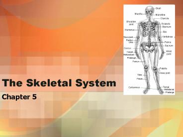
The Skeletal System
Description:
Junqueira and Carneiro, Basic Histology, a text and atlas, pp. 44 and 46, Figures 8-6 and 8-8. ... Tibial tuberosity: where ligaments of patella attach ... – PowerPoint PPT presentation
Number of Views:35
Avg rating:3.0/5.0
Title: The Skeletal System
1
The Skeletal System
- Chapter 5
2
Long bone
3
Haversian System
Junqueira and Carneiro, Basic Histology, a text
and atlas, pp. 44 and 46, Figures 8-6 and 8-8.
4
Bone growth
Epiphyseal plates
5
Axial Skeleton
- 3 parts
- 1. Skull
- 2. vertebral column
- 3. bony thorax
- The skull consists of
- -cranium (around brain)
- -facial bones
6
Cranium
- 8 bones
- Frontal (forehead)
- Parietal (paired laterally)
- Temporal (paired laterally)
- Occipital (posterior cranium)
- Sphenoid (butterfly shaped part of brain base
and behind eye orbit) - Ethmoid (roof of nasal cavity)
7
Cranium
- Coronal suture where frontal and parietal bones
meet - Sagittal suture where parietal bones meet
- Squamous suture where temporal and parietal
meet - Lambdoidal suture junction of occipital with
parietal bones
8
(No Transcript)
9
(No Transcript)
10
(No Transcript)
11
(No Transcript)
12
Facial Bones
13
Fetal skull
- Fontanels fibrous membranes connecting cranial
bones soft spots - For brain growth and skull compression during
birth - Converted to bone by 22-24 months
14
Vertebral column
15
Pectoral Girdle
- Clavicle collarbone
- Scapula shoulder blades
- Glenoid cavity of scapula is where humerus fits
Scapula
16
Upper Limbs
Head of humerus (glenoid fossa)
Radial groove for nerve (seen on back
side) Deltoid tuberosity
humerus
capitulum
epicondyle
Radial tuberosity
radius
trochlea
ulna
ulna
(a)
(b)
Anterior view, left arm (a) anatomical
position (b) hand pronated
Image www.tpub.com/.../medical/14295/css/14295_23
.htm
17
Image from Wikimedia
18
The hand
- Carpals wrist
- Metacarpals palm
- Phalanges fingers
Distal end of radius is always on thumb side.
Image www.heumann.org/.../appendicular.skeleton.h
tml
19
Pelvic Girdle
- Formed from 2 coxal (hip) bones
- Pelvis coxal bones sacrum coccyx
- Bears weight of upper body
- Protects reproductive, urinary organs, and
intestines
False pelvis
True pelvis
Image www.heumann.org/.../appendicular.skeleton.h
tml
20
Pelvis
- Female pelvis (vs. male)
- Shallower, thinner --shorter sacrum
- Ilia flare laterally --larger inlet of true
pelvid - Rounded pubic arch
Male female
21
Lower limbs
Femur Slants medially to put knees in line with
center of gravity
22
Lower limbs
- Tibia (shinbone)
- Larger then fibula
- Tibial tuberosity where ligaments of patella
attach - Medial malleolus inner ankle (on big toe side)
- Fibula
- Lateral malleolus outer ankle
23
Foot
heel
ankle
24
Joints (articulations)
- Hold bones together, yet provide for movement
- Functional classification
- Synarthroses immovable
- Amphiarthroses slightly movable
- Diarthroses freely movable
- Structural classification
- Fibrous (generally synarthroses)
- Cartilagenous (mostly amphiarthroses)
- Synovial (diarthroses)
25
Structural Joints
- Fibrous
- In sutures of skull
- Bound by connective tissue fibers
- Cartilagenous
- Bone ends covered by cartilage
- Pubic symphysis, invertebral joints (slightly
movable) - Epiphyseal plates, between 1st ribs and sternum
(immovable)
26
Structural Joints
- Synovial
- Connected by joint cavity with synovial fluid
- 4 key features
- Hyaline cartilage at bone ends
- Capsule of fibrous connective tissue lined with
synovial membrane - Joint cavity with synovial fluid
- Reinforcing ligaments
- Bursae and tendon sheaths help reduce friction at
muscles/ligaments/tendons
27
Types of synovial joints
Mulitaxial, rotation
Rotating uniaxial
Uniaxial angular
28
Types of synovial joints
Biaxial, side to side Back and forth
A.k.a. plane joint Flat w/ no rotation
Biaxial (like condyloid) movement
Image www.visualdictionaryonline.com
29
(No Transcript)


