ENDOCRINE SYSTEM ENDOCRINE GLANDS - PowerPoint PPT Presentation
1 / 31
Title:
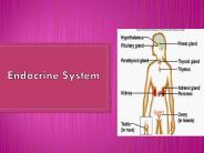
ENDOCRINE SYSTEM ENDOCRINE GLANDS
Description:
endocrine gland: hypophysis, thyroid gland, parathyroid gland, ... triangle or semilunar shape. 8-20g, 4-6cm length, 1-2cm width, 0.5cm thickness. capsule ... – PowerPoint PPT presentation
Number of Views:350
Avg rating:3.0/5.0
Title: ENDOCRINE SYSTEM ENDOCRINE GLANDS
1
ENDOCRINE SYSTEM(ENDOCRINE GLANDS)
2
- nervous system
- electrochemical signals
- endocrine system
- chemical agents hormone
- characteristics of the endocrine gland
- ductless
- well vascularized
- hormone secretion
3
- endocrine gland hypophysis, thyroid gland,
parathyroid gland, adrenal gland, pineal body,
pancreas, testis, ovary, liver
4
I. HYPOPHYSIS
- 1 x 1.5 x 0.5 cm, 0.5 gr
- master gland located in sella trucica
5
(No Transcript)
6
- oral ectodermal origin adenohypophysis
- pars distalis, pars tuberalis, pars intermedia
- neural ectodermal origin neurohypophysis
- pars infundibularis, pars nervosa
- anterior and posterior lobe
- anterior lobe pars distalis, pars tuberalis
- posterior lobe pars nervosa, pars intermedia
7
- 1. Adenohypophysis
- 1) Pars distalis
- 75 of total hypophysis
- Chromophils(50) 12-15µm
- acidophils(a-cell 35) basophils(ß-cell)
- Chromophobe(50)
- smaller than chromophils
8
- A. Acidophils
- somatotroph growth hormone
- mammotroph(lactotroph) prolactin
- B. Basophils
- gonadotroph follicular stimulating hormone(FSH)
- luteinizing hormone(LH) or
- interstitial cell stimulating hormone(ICSH)
- thyrotroph thyroid stimulating hormone(TSH)
- corticotroph adrenocorticotrophic hormone(ACTH)
- C. Chromophobes
- stem cell
- inactivated chromophils
9
- 2) Pars intermedia
- 3 of the human fetal hypophysis
- degeneration in adults
- well developed in some animals
- especially in amphibians MSH secretion
- 3) Pars tuberalis
- Chromophobes, chromophils(gonadotropins)
10
- 2. Neurohypophysis
- axon and axon terminal from hypothalamus
hypothalamo-hypophyseal tract - neuronal cell body in nucleus of the
hypothalamus - suprachiasmatic and paraventricular nucleus
- release oxytocine, vasopressin
- dorsal medial N, ventral medial N, and
infundibular N - release neuropeptide to the adenohypophysis
- via the portal system
- neuroglial cells(pituicyte)
11
- 3. Blood vessels
- internal carotid a ? superior and inferior
hypophyseal artery - superior hypophyseal a.
- ? pars infundibularis
- ? primary capillary plexus
- ? hypophyseal portal vein
- ? adenohypophysis
- ? secondary capillary plexus
- ? vein ? cavernosus sinus
- inferior hypophyseal a.
- ? pars nervosa
- ? capillary plexus
- ? vein ? cavernosus sinus
12
4. Hypophysis and hypothalamus hypophyseal
portal system and hypothalamohypophyseal tract
13
(No Transcript)
14
II. Adrenal gland(Suprarenal gland)
- paired organs
- located in superior pole of each kidney
- triangle or semilunar shape
- 8-20g, 4-6cm length,
- 1-2cm width, 0.5cm thickness
- capsule
- cortex mesordermal origin(80-90)
- medulla neural crest orgin(10-20)
15
(No Transcript)
16
- 1. Adrenal cortex
- 3 layers glomerulosa, fasciculata, reticularis
- (1) Zona glomerulosa
- 15 of cortex
- columnar or pyramidal cells round or ellipsoisal
arrangement - mineralocorticoids aldosterone
- (2) Zona fasciculata
- 78 of cortical volume
- polyheadral cells longitudinal column
- glucocorticoids cortisol, corticosterone
- small amount of androgen and estrogen
- (3) Zona reticularis
17
2. Adrenal medulla large epitheloid cells in
cluster or cell cords chromaffin
cells norepinephrine, epinephrine nerve cell
18
(No Transcript)
19
- 3. Blood supply of the adrenal gland
- superior(from inferior phrenic artery),
middle(from aorta) and inferior(from renal
artery) suprarenal artery - ? subcapsular arterial plexus(SAP)
20
i, SAP ? short cortical artery(cortical atery) ?
sinusoid(in cortex) ? venule(in medulla) ?
suprarenal vein ii, SAP ? long cortical
artery(medullary artery) ?capillary network(in
medulla) ? venule ? suprarenal vein
21
- 4. Histophysiology of the adrenal cortex
- (1) Hyperadrenocorticism
- Cushings syndrome
- excessive secretion of ACTH
- excessive secretion of glucocorticoids
obesity(face, neck, trunk), hirsutisim,
impotence, amenorrhea - Conn's syndrome
- excessive secretion of aldosterone
- (2) Hypoadrenocorticism(Addison's disease)
- destruction of adrenal cortex
- weakness, weight loss, low blood pressure,
increase skin pigmentation
22
III. Thyroid gland
- 1. Anatomical Structure
- 25-40g
- thyroid cartilage
- 6th tracheal cartilage
- two lateral lobe
- isthmus
- pyramidal lobe
23
2. Blood Supply
24
- 3. Histological organization
- colloid follicle
- follicular(principal) cell
- thyroxin
- parafollicular cell
- calcitonin
Fig. 15-43. Thyroid gland. H-E stain
25
- 4. Histophysiology of the thyroid gland
- (1) Thyroid hormone
- increase the basal metabolic rate
- absorption of carbohydrates
- protein synthesis development, differentiation,
growth - a. hypothyroidism
- lowering metabolic rate
- in child
- Cretinism mental retardation, dwarfing
- in adults myxedema
- b. hyperthyroidism(thyrotoxicosis)
- Grave's disease or exopthalmic goiter
- c. thyroid stimulating hormone
- (2) Calcitonin
26
IV. Parathyroid gland
- 4-6 glands
- located on posterior surface of the thyroid
gland - 5mm in length, 4mm in width, 2mm in thickness
- 25-50mg
27
cell groups chief cell parathyroid
hormone oxyphil cell unknown function
28
- Histophysiology of the parathyroid gland
- parathyroid hormone(PTH)
- increase blood calcium level
- Hyperparathyroidism
- decrease blood phosphate level, increase blood
calcium level - pathological deposition of Ca soft tissue
calcification, bone fracture - Hypoparathyroidism
- decrease blood calcium level, increase blood
phosphate level - over excitability of nervous system, tetany
29
V. Pineal body pineal gland, epiphysis cerebri
- posterior end of roof of the 3rd ventricle
- 5-8mm in length, 3-5mm in width
- 0.1-0.2 g
- Cells
- pinealocyte melatonin secretion
- interstitial cell astrocyte like cell
30
- Corpora arenacea(brain sand)
- calcium phosphate, carbonate
- land mark for X-ray, CT
31
- Histophysiology of the pineal gland(continue)
- seaonal changes in melatonin and testes
- Syrian hamster
- in summer 3,000 mg weight of testes
- in winter 300 mg
- pineal gland, melatonin and adolescence
- pineal peptides arginin vasotocin,
angigonadotrophin, gonadotrophin releasing factor - neuroepiphysins
- Descartes "seat of the rational soul
- seasonal mood disorsers, jet lag
Recommended
CrystalGraphics Presentations