CLS Status Update - PowerPoint PPT Presentation
Title:
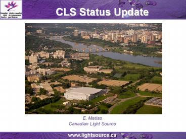
CLS Status Update
Description:
CLS Status Update E. Matias Canadian Light Source – PowerPoint PPT presentation
Number of Views:93
Avg rating:3.0/5.0
Title: CLS Status Update
1
CLS Status Update
E. Matias Canadian Light Source
2
Layout
170.88 m circumference 2.9 GeV 225-300
mADBA lattice with 12-fold period
3
Major Facility Projects
- Complete Phase II Beamlines
- Start Phase III Beamlines
- New Linac RF System
- New Linac Transfer Line Power Supplies
4
Phase II - Under Construction
Beamline (Port) Energy (eV) Energy (eV) Resolution (?E/E) Flux (?/s/0.1BW) _at_ 500 mA Techniques
Beamline (Port) Wavelength (Å) Wavelength (Å) Resolution (?E/E) Spot size Hor x Vert Techniques
Resonant Elastic and Inelastic X-ray Scattering (REIXS)1 (10ID-2) 80 2000 1 x 10⁻⁴ _at_100 eV 2 x 10⁻⁴ _at_1000 eV 2 x 10¹³ _at_ 100 eV 5 x 10¹² _at_ 1000eV 250 µm x 15 µm 60 µm x 10 µm X-ray Absorption Spectroscopy X-ray Emission Spectroscopy Resonant Inelastic X-ray Scattering Resonant Elastic X-ray Scattering Coherent X-ray Scattering (Speckle) Magnetic X-ray Dichroism Molecular Beam Epitaxy sample preparation
Resonant Elastic and Inelastic X-ray Scattering (REIXS)1 (10ID-2) 155 6.2 1 x 10⁻⁴ _at_100 eV 2 x 10⁻⁴ _at_1000 eV 2 x 10¹³ _at_ 100 eV 5 x 10¹² _at_ 1000eV 250 µm x 15 µm 60 µm x 10 µm X-ray Absorption Spectroscopy X-ray Emission Spectroscopy Resonant Inelastic X-ray Scattering Resonant Elastic X-ray Scattering Coherent X-ray Scattering (Speckle) Magnetic X-ray Dichroism Molecular Beam Epitaxy sample preparation
Soft X-ray Microcharacterization Beamline (SXRMB)1 (06B1-1) 1700 10000 3.3 x 10⁻⁴ InSb (III) 1 x 10⁻⁴ InSb (III) XAFS gt 1 x 10¹¹ Microprobe 1 x 10⁹ 300 µm x 300 µm 10 µm x 10 µm X-ray Absorption Spectroscopy Microprobe X-ray Excited Optical Luminescence (XEOL) Resonant spectroscopies X-ray Magnetic Linear Dichroism (XMLD) Photo Emission Electron Microscopy (PEEM) X-ray Magnetic Circular Dichroism (XMCD) Photo and Auger Electron Spectroscopy
Soft X-ray Microcharacterization Beamline (SXRMB)1 (06B1-1) 7.3 1.3 3.3 x 10⁻⁴ InSb (III) 1 x 10⁻⁴ InSb (III) XAFS gt 1 x 10¹¹ Microprobe 1 x 10⁹ 300 µm x 300 µm 10 µm x 10 µm X-ray Absorption Spectroscopy Microprobe X-ray Excited Optical Luminescence (XEOL) Resonant spectroscopies X-ray Magnetic Linear Dichroism (XMLD) Photo Emission Electron Microscopy (PEEM) X-ray Magnetic Circular Dichroism (XMCD) Photo and Auger Electron Spectroscopy
Synchrotron Laboratory for Micro And Nano Devices (SyLMAND)1 (03B1-1 03B2-1) 1000 15000 NA NA 150 mm x 15 mm Deep X-ray lithography LIGA process lithography steps
Synchrotron Laboratory for Micro And Nano Devices (SyLMAND)1 (03B1-1 03B2-1) 12.4 0.82 NA NA 150 mm x 15 mm Deep X-ray lithography LIGA process lithography steps
Very Sensitive Elemental and Structural Probe Employing Radiation from a Synchrotron (VESPERS)1 (07B2-1) 6000 30000 Si(111) 10-4 MLM1 10-2 MLM2 10-1 Pink Beam Si-111 2 x 10⁹ _at_ 15 keV MLM1 1 x 10¹¹ _at_ 15 keV MLM2 4 x 10¹¹ _at_ 15 keV (2 4) µmx(2\ 4) µm X-ray Laue Diffraction X-ray Fluorescence Spectroscopy X-ray Absorption Near Edge Structure Differential Aperture X-ray Microscopy Multi-bandpass and pink beam capability
Very Sensitive Elemental and Structural Probe Employing Radiation from a Synchrotron (VESPERS)1 (07B2-1) 2 0.4 Si(111) 10-4 MLM1 10-2 MLM2 10-1 Pink Beam Si-111 2 x 10⁹ _at_ 15 keV MLM1 1 x 10¹¹ _at_ 15 keV MLM2 4 x 10¹¹ _at_ 15 keV (2 4) µmx(2\ 4) µm X-ray Laue Diffraction X-ray Fluorescence Spectroscopy X-ray Absorption Near Edge Structure Differential Aperture X-ray Microscopy Multi-bandpass and pink beam capability
Canadian Macromolecular Crystallography Facility (CMCF 2)1 (08B1-1) 4000 18000 10-4 1012 _at_ 12 keV 230 µm x 160 µm X-ray diffraction Multiwavelength Anomalous Dispersion (MAD)
Canadian Macromolecular Crystallography Facility (CMCF 2)1 (08B1-1) 3.1 0.69 10-4 1012 _at_ 12 keV 230 µm x 160 µm X-ray diffraction Multiwavelength Anomalous Dispersion (MAD)
Biomedical Imaging and Therapy (BMIT - BM)1 (05B1-1) 8000 40000 M1 DEI 10-4 1.5 x 10¹³ _at_ 10 keV 231 mm x 4.6 mm _at_ 23 m Conventional absorption imaging Diffraction Enhanced Imaging (DEI) / Multiple Image Radiography (MIR) Phase contrast or in-line holography Ultra-small, small, wide angle scattering imaging Computer Tomography (CT)
Biomedical Imaging and Therapy (BMIT - BM)1 (05B1-1) 1.6 0.3 M1 DEI 10-4 1.5 x 10¹³ _at_ 10 keV 231 mm x 4.6 mm _at_ 23 m Conventional absorption imaging Diffraction Enhanced Imaging (DEI) / Multiple Image Radiography (MIR) Phase contrast or in-line holography Ultra-small, small, wide angle scattering imaging Computer Tomography (CT)
Biomedical Imaging and Therapy (BMIT - ID)1 (05ID-1 05ID-2) 20000 100000 M1 CT10-3 M2 CT10-3 M3 DEI105 M4 KES10-3 4 x 10¹⁴ _at_ 40 keV 224 mm x 11.2 mm _at_ 56 m Imaging conventional absorption imaging, DEI / MIR, CT, K-edge Subtraction (KES) Therapy Microbeam Radiation Therapy, CT Therapy
Biomedical Imaging and Therapy (BMIT - ID)1 (05ID-1 05ID-2) 0.6 0.1 M1 CT10-3 M2 CT10-3 M3 DEI105 M4 KES10-3 4 x 10¹⁴ _at_ 40 keV 224 mm x 11.2 mm _at_ 56 m Imaging conventional absorption imaging, DEI / MIR, CT, K-edge Subtraction (KES) Therapy Microbeam Radiation Therapy, CT Therapy
B1/B2 - Indicates Bending Magnet Beamline 1
- operational in 2008 ID - Indicates
Insertion Device Beamline
5
BMIT Licensing
- Controls Related
- Safety System Design (Based on IEC 61508)
- Human FactorsEngineering Review
6
Phase III
The Brockhouse X-ray Diffraction and Scattering Sector (Undulator)2 (04ID-1) 3000 15000 Inelastic Scattering 10-6 1010 Into 100 µm X 100 µm Inelastic Scattering Resonant Scattering Grazing Incidence Diffraction Reflectivity Small Angle X-ray Scattering Wide Angle X-ray Scattering
The Brockhouse X-ray Diffraction and Scattering Sector (Undulator)2 (04ID-1) 3000 15000 Resonant Scattering 10-4 1012 Into 100 µm X 100 µm Inelastic Scattering Resonant Scattering Grazing Incidence Diffraction Reflectivity Small Angle X-ray Scattering Wide Angle X-ray Scattering
The Brockhouse X-ray Diffraction and Scattering Sector (Undulator)2 (04ID-1) 3000 15000 Grazing Incidence Diffraction 10-4 1012 Into 100 µm X 200 µm Inelastic Scattering Resonant Scattering Grazing Incidence Diffraction Reflectivity Small Angle X-ray Scattering Wide Angle X-ray Scattering
The Brockhouse X-ray Diffraction and Scattering Sector (Undulator)2 (04ID-1) 4.1 0.8 Grazing Incidence Diffraction 10-4 1012 Into 100 µm X 200 µm Inelastic Scattering Resonant Scattering Grazing Incidence Diffraction Reflectivity Small Angle X-ray Scattering Wide Angle X-ray Scattering
The Brockhouse X-ray Diffraction and Scattering Sector (Undulator)2 (04ID-1) 4.1 0.8 SAXS/WAXS10-4 1011 Into 200 µm X 100 µm Inelastic Scattering Resonant Scattering Grazing Incidence Diffraction Reflectivity Small Angle X-ray Scattering Wide Angle X-ray Scattering
The Brockhouse X-ray Diffraction and Scattering Sector (Wiggler)2 (04ID-2) 7000 60000 Powder diffraction10-4 1011 Into 1000 µm X 200 µm Powder Diffraction Single Crystal Crystallography High Pressure Diamond Anvil Cell High Q-space PDF
The Brockhouse X-ray Diffraction and Scattering Sector (Wiggler)2 (04ID-2) 7000 60000 Single crystal crystallography10-3 1010 Into 100 µm X 100 µm Powder Diffraction Single Crystal Crystallography High Pressure Diamond Anvil Cell High Q-space PDF
The Brockhouse X-ray Diffraction and Scattering Sector (Wiggler)2 (04ID-2) 1.8 0.2 High pressure diamond anvil10-4 1011 Into 25 µm X 25 µm Powder Diffraction Single Crystal Crystallography High Pressure Diamond Anvil Cell High Q-space PDF
The Brockhouse X-ray Diffraction and Scattering Sector (Wiggler)2 (04ID-2) 1.8 0.2 High Q space PDF10-3 1011 Into 500 µm X 500 µm Powder Diffraction Single Crystal Crystallography High Pressure Diamond Anvil Cell High Q-space PDF
BioXAS Life Science Beamline for X-ray Absorption Spectroscopy (Wiggler)2 (07ID-1) 5000 28000 10-4 9x10¹² into 600 µm X 200 µm Bulk X-ray Absorption Fine Structure at low and ultra-low concentrations and high resolution with automation for FedEXAFS XAS Imaging Macro (gt50 mm), Micro (2 mm), and Sub-Micro (lt1 to 2mm)
BioXAS Life Science Beamline for X-ray Absorption Spectroscopy (Wiggler)2 (07ID-1) 2.5 0.4 10-4 9x10¹² into 600 µm X 200 µm Bulk X-ray Absorption Fine Structure at low and ultra-low concentrations and high resolution with automation for FedEXAFS XAS Imaging Macro (gt50 mm), Micro (2 mm), and Sub-Micro (lt1 to 2mm)
BioXAS Life Science Beamline for X-ray Absorption Spectroscopy (Undulator)2 (07ID-2) 5000 15000 10-4 2x10¹³ into 400 µm X 20 µm XAS Imaging Macro (gt50 mm), Micro (2 mm), and Sub-Micro (lt1 to 2mm)
BioXAS Life Science Beamline for X-ray Absorption Spectroscopy (Undulator)2 (07ID-2) 2.5 0.8 10-4 2x10¹³ into 400 µm X 20 µm XAS Imaging Macro (gt50 mm), Micro (2 mm), and Sub-Micro (lt1 to 2mm)
The Quantum Materials Spectroscopy Centre (EPU1 225 mm period)2 (09ID-1) 10 200 NA Brilliance 7 x 10¹⁶ Spin 400 µm X 40 µm 400 µm X 40 µm (at 30 eV) Spin and angle resolved photoemission spectroscopy Separate organic and inorganic Molecular Beam Epitaxy and characterization chambers
The Quantum Materials Spectroscopy Centre (EPU1 225 mm period)2 (09ID-1) 1240 62 NA Brilliance 7 x 10¹⁶ Spin 400 µm X 40 µm 400 µm X 40 µm (at 30 eV) Spin and angle resolved photoemission spectroscopy Separate organic and inorganic Molecular Beam Epitaxy and characterization chambers
The Quantum Materials Spectroscopy Centre (EPU2 54 mm period)2 (09ID-2) 200 1000 NA Brilliance 7 x 10¹⁷ Spin 700 µm X 40 µm ARPES 300 µm X 40 µm (at 200 eV) Spin and angle resolved photoemission spectroscopy Separate organic and inorganic Molecular Beam Epitaxy and characterization chambers
The Quantum Materials Spectroscopy Centre (EPU2 54 mm period)2 (09ID-2) 62 12 NA Brilliance 7 x 10¹⁷ Spin 700 µm X 40 µm ARPES 300 µm X 40 µm (at 200 eV) Spin and angle resolved photoemission spectroscopy Separate organic and inorganic Molecular Beam Epitaxy and characterization chambers
B1/B2 - Indicates Bending Magnet Beamline
ID - Indicates Insertion Device
Beamline 2 CFI funded not yet approved
for construction.
7
EPICS Environment
- IOC Hardware
- Motorola 68360 Single board computers
(approximately 150) - Moxa IOCs (approximately 50)
- VME 64x with SIS Optical Links (approximately
25-30) - CosyLab MicroIOC (3)
- Many Server Based Soft IOCs.
- IOC Software
- Scientific Linux or RTEMS
- EPICS 3.14.6
- High level Software
- EDM,
- Matlab, ROOT, assorted packages
- Web Services (next week)
- PLC
- Modicon Momentum (approximately 40)
- Siemens S7/300 (4)
- Siemens S7/400 (2)
8
Controls and Instrumentation Development Specific
Projects
- Time-resolve/Single Bunch
- Fill-pattern monitor (J. Vogt/J. Bergstrom)
- EPICS Software (D. Beauregard)
- Transverse Feedback System (S. Hu)
- Upgraded timing system
- Alarm Annunciation (D. Beauregard)
- Beam profile analysis
- Beamline Java Interface ..
- ScienceStudio/Remote Access ..
9
Fill Pattern Monitor
- Complete Phase II Beamlines
- Start Phase III Beamlines
- New Linac RF System
- New Linac Power Supplies
10
Results.
11
Results.
12
Audio Alarm (using Festival)
13
Virtual PA System
14
Funding Partners
38 supporting University Partners and growing


