Respiratory Slide Show - PowerPoint PPT Presentation
1 / 25
Title:
Respiratory Slide Show
Description:
3. Cricoid cartilage. 4. Interarytenoid muscle. Formation of the Larynx. 1. Vestibule ... Formation of Lower Respiratory Tract: The Role of Induction ... – PowerPoint PPT presentation
Number of Views:42
Avg rating:3.0/5.0
Title: Respiratory Slide Show
1
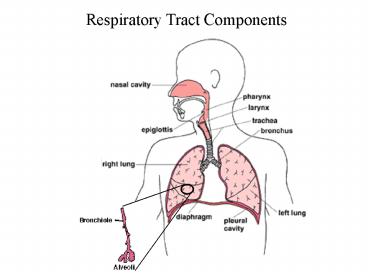
Respiratory Tract Components
2
Respiratory Tract Components in the Bird
lung
3
Path of Air into Lungs
Nostrils Nasal Cavity Nasopharynx Larynx
4
Formation of the Larynx
5
Formation of the Larynx
6
Formation of the Larynx
1. Thyroid cartilage 2. Arytenoid cartilage 3.
Cricoid cartilage 4. Interarytenoid muscle
7
Formation of the Larynx
1. Vestibule 2. False vocal cord 3. True
vocal cord 4. Trachea
8
Path of Air into Lungs
Nostrils Nasal Cavity Nasopharynx Larynx
Trachea Bronchi Bronchioles Alveolii
9
Formation of Upper Respiratory Tract
10
Lower Respiratory Tract
11
Formation of Lower Respiratory Tract
12
Formation of Lower Respiratory Tract
13
Formation of the Laryngotracheal Tube
14
Formation of Lower Respiratory Tract
tracheoesophageal septum
15
Formation of the Larynx
16
Formation of Lower Respiratory Tract
Recanalization of Larynx
17
Formation of Lower Respiratory Tract
Why are some tubes straight and others branched?
18
Formation of Lower Respiratory Tract The Role of
Induction
Gut Mesenchyme
Thoracic Mesenchyme
19
Formation of Lower Respiratory Tract The Role of
Induction
20
Formation of Lower Respiratory Tract The Role of
Induction
21
Formation of Lower Respiratory Tract
22
Development of Lungs and the Pleura Cavity
23
Development of Lungs and the Pleura Cavity
Pseudoglandular phase (5-17 weeks) Further
branching of the duct system (up to 21 further
orders) generates the presumptive conducting
portion of the respiratory system up to the
level of the terminal bronchioles. At this time
the future airways are narrow with little lumens
and a pseudostratified squamous epithelium. They
are embedded within a rapidly proliferating
mesenchyme. The structure has a glandular
appearance. Canalicular phase (15-25
weeks) The onset of this phase is marked by
extensive angiogenisis within the mesenchyme that
surrounds the more distal reaches of the
embryonic respiratory system to form a dense
capillary network. The diameter of the airways
increases with a consequent decrease in
epithelial thickness to a more cuboidal
structure. The terminal bronchioles branch to
form several orders of respiratory bronchioles.
Differentiation of the mesenchyme progresses
down the developing respiratory tree, giving rise
to chondrocytes, fibroblasts and myoblasts.
Terminal sac phase (24-40weeks) Branching
and growth of the terminal sacs or primitive
alveolar ducts. Continued thinning of the stroma
brings the capillaries into apposition with the
prospective alveoli. The prealveoli cells then
flatten, increasing the epithelial surface area
by dilation of the saccules, giving rise to
immature alveoli. By 26 weeks, a rudimentary
though functional blood/gas barrier has formed.
Maturation of the alveoli continues by further
enlargement of the terminal sacs, deposition of
elastin foci and development of vascularised
septae around these foci. The stroma continues
to thin until the capillaries protrude into the
alveolar spaces. Alveolar phase (36 weeks
- term/adult) Maturation of the lung
indicated by the appearance of fully mature
alveoli begins at 36 weeks, though new alveoli
will continue to form for approximately three
years. A decrease in the relative proportion of
parenchyma to total lung volume still
contributes significantly to growth for 1 to 2
years after birth, thereafter all components
grow proportionately until adulthood.
24
(No Transcript)
25
Pulmonary Vasculature


