The Brain - PowerPoint PPT Presentation
1 / 37
Title:
The Brain
Description:
Pons: sleep & arousal, attention, assists in movement. Medulla: unconscious vital functions like ... Reticular Activating System: wakefulness & sleep, alertness ... – PowerPoint PPT presentation
Number of Views:63
Avg rating:3.0/5.0
Title: The Brain
1
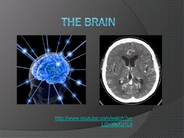
The Brain
- http//www.youtube.com/watch?vLi5nMsXg1Lk
2
- Which brain imaging technique injects a substance
in order to view active areas of the brain
because of glucose consumption? - Which brain imaging technique graphs brain waves
and is mostly used for sleep research? - Which brain imaging technique combines
cross-sectional x-rays to view the structure of
the brain?
3
- Too little of this neurotransmitter is associated
with Parkinsons? - Too little of this neurotransmitter is associated
with anxiety disorders? - Too little of this neurotransmitter is associated
with Alzheimers - Too little of this neurotransmitter is associated
with depression? - Too much of this neurotransmitter is associated
with schizophrenia?
4
(No Transcript)
5
Hindbrain
- Pons sleep dreaming, arousal, attention,
assists in movement - Medulla unconscious vital functions like
breathing, circulation, etc. - Reticular Activating System wakefulness sleep,
alertness
6
Hindbrain
- Cerebellum balance, motor coordination
7
Midbrain
- Integrates types of sensory info and muscle
movements
8
Limbic System
- Thalamus
- Relays sensory info from the body to parts of
the brain - Hypothalamus
- Maintains homeostasis regulates body
temperature, hunger, thirst, blood pressure,
hormones, etc.
9
Limbic System
- Amygdala
- Emotional responses, particularly aggression
attention to novel stimuli - Hippocampus
- Formation of memories
- Pituitary Gland
- Master gland secretes hormones
10
Which part of the brain?
? Pons
- REM sleep dreaming, assists in movement
- Relay station for sensory info
- On switch for the brain, alertness
wakefulness, attention - Body temperature, hunger, thirst, glands
- Balance and motor coordination
- Emotions (aggression), novel stimuli
- Master gland
- Unconscious essential functions such as
respiration and heart rate - Formation of new memories
? Thalamus
? R.A.S.
?Hypothal.
? Cerebellum
? Amygdala
? Pituitary gland
? Medulla
? Hippocampus
11
Brain Lateralization
- Cerebrum
- Surface of brain, two hemispheres, thinking
language - Cerebral Cortex
- Wrinkled, convoluted surface divided into
four lobes - Corpus Callosum
- Thick bundle of fibers which connects the two
hemispheres
12
Hemispheres of the Brain
13
Left Hemisphere Rational, Logical
- Language
- Math
- Responds to verbal instructions
- Right side of body
14
Right Hemisphere Intuitive, Artistic
- Visual imagery
- Music
- Spatial abilities
- Responds to demonstrated instructions
- Left side of the body
15
(No Transcript)
16
(No Transcript)
17
(No Transcript)
18
(No Transcript)
19
Lobes of the Brain
20
Frontal Lobe
- Responsible for abstract thought, emotion,
voluntary body movements, language
21
Parietal Lobe
- Receives incoming touch, pressure, and pain
sensations from the body
22
Occipital Lobe
- Located at the rear of the brain
- Involved in the reception and interpretation of
visual information
23
Temporal Lobe
- Located on the side, slightly above ears
- Involved in reception and interpretation of
auditory stimuli
24
Language the Brain
- Brocas Area
- Physical production of speech
- Brocas Aphasia
- Inability to physically speak words
25
Language the Brain
- Wernickes Area
- Comprehension of language
- Wernickes Aphasia
- Inability to understand language/words
- Word salad
26
- http//www.ted.com/talks/lang/eng/jill_bolte_taylo
r_s_powerful_stroke_of_insight.html
27
The Split-Brain Experiments
- 1960s, Roger Sperry
- Epilepsy seizures spread to other hemisphere
through corpus callosum - In his operations, the entire corpus callosum was
severed hemispheres completely independent of
one another - "The great pleasure and feeling in my right
brain is more than my left brain can find the
words to tell you. Roger Sperry
28
The Split-Brain Experiments
- Michael Gazzaniga more experiments
- Patients appeared normal (talk, read, alert,
etc.) - BUTif patient held up something like coffee cup
in left hand, couldnt speak its name - If object in right hand, no trouble at all
- Printed word LOUSE visible only in left visual
field, couldnt read ? put in right side, could
read it fine
Right vision field is connected to the left
hemisphere. Left vision field is connected to the
right hemisphere.
29
Split-Brain Operations
- Only sever portion of corpus callosum (splenium
remains intact) - Split brain patients learn very quickly how to
keep both sides in communication
30
Phineas Gage
31
Lesioning
- Studying damaged parts of the brain to observe
the changes in behavior or loss of function
32
Ways of Studying the Brain
- Electroencephalogram (EEG) detects and records
brain-wave activity - Widely used in sleep research
33
- Computerized Axial Tomography (CAT) scan uses
X-ray cameras that rotate around the brain and
combine pictures into a detailed image of the
brains structure - Does not give info about function
34
- Positron Emission Tomography (PET) scan measures
how much of a chemical (i.e. glucose or oxygen)
that parts of the brain are using, which
indicates activity - Indicates function, but expensive
35
- Magnetic Resonance Imaging (MRI) similar to CAT
scan but produces higher-resolution images - Patient lies in magnetic field is exposed to
radio waves that record activity of brain due to
blood flow
36
- Functional MRI (fMRI) combines MRI and PET
scanning techniques, detects metabolic (i.e.
changes in blood flow) in parts of brain - Used for brain mapping
37
Magnetoencephalogram (MEG)
- Measures the very faint magnetic fields that are
emitted from the head as a result of brain
activity - Magnetic detection coils are bathed in liquid
helium held over persons head brains
magnetic field induces current in the coils - Induces a magnetic field using an instrument
known as a superconducting quantum interference
device (SQUID)


