Cantilevers - PowerPoint PPT Presentation
1 / 25
Title:
Cantilevers
Description:
The cantilever elastic costant k must allow not negligible deflection under ... The ceramic is heated above its Curie temperature Tc( 200 C), and then it is ... – PowerPoint PPT presentation
Number of Views:342
Avg rating:3.0/5.0
Title: Cantilevers
1
Cantilevers
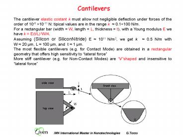
The cantilever elastic costant k must allow not
negligible deflection under forces of the order
of 10-8 10-11 N tipical values are in the range
k 0.1100 N/m. For a rectangular bar (width
W, length L, thickness t), with a Young
modulus E we have k E(t/L)3W/4. Assuming
(Silicon or SiliconNitride) E 1011 N/m2, we get
k 0.5 N/m with W 20 µm, L 100 µm, and
t 1 µm. The most flexible cantilevers (e.g.
for Contact Mode) are obtained in a rectangular
geometry that offers high sensitivity to lateral
force More stiff cantilever (e.g. for
Non-Contact Modes) are Vshaped and insensitive
to lateral force
2
Cantilevers (2)
The mechanical resonant frequency fr of a
cantilever with one fixed end is proportional to
v(E/?)t/L2, where ? is the density and E the
Young modulus. To increase fr while keeping
constant the stiffness (k), the cantilever must
be compact. High resonant frequency allows
faster scans tipically the maximum scan rate fs
(1/Ts, where Ts is the time interval between
two signal acquisitions) is chosen fs fr/100.
Commercial cantilevers offer fr ranging from a
few kHz to fraction of MHz.
3
Tips
Different commercial tips are available with
different values of the apex curvature radius R
typically Pyramidal R 500 Å Sharpened
pyramidal R 200 Å Conical (ultratip)
R 50100 Å
Pyramidal tips are obtained by photolithography
through a squared hole in a mask evaporated onto
a silicon wafer, a pyramidal pit is produced by
anisotropic etching of 001 plane then a
SiliconNitride film is deposited and glued onto a
glass plate. By chemical etching the silicon is
removed leaving the SiliconNitride cantilever
with pyramidal tip. Conical tip are grown by SEM
deposition in dirty atmosphere, or ion-milling
(FIB Focused Ion Beam)
4
Aspect Ratio and Curvature Radius
An important parameter is the aspect ratio (ratio
between tip height and base) The larger is the
aspect ratio the better is resolved the SPM
image. Alternatively the tip quality is defined
by the sidewall angle, that for a pyramidal tip
is 45 or 62.5 (edge or face of the tetrahedron
) and for conical tips varies from base to apex
reaching a maximum of about 70.
FIB tips
Very high Aspect Ratio tips may be obtained from
nanotubes
5
Nanotube tips high aspect ratio
6
Sources of Image artifacts
Z offset - Tilt - Non-linearity Drift - Blur -
Glitches
7
Scanner non-ideality
Typical scanner imperfections are Non-linearity C
reep Hysteresis Aging Thermal drift
Cross-coupling among x-y-z displacements
Non-linearity hysteresis
Creep
Scanner non-linearity introduces image
distortions to improve linearity secondary
position detectors (optical, capacitive) may be
used, or a correction through non-linear
transfer function (LUT Look-Up-Table) may be
calculated through scanner calibration, measuring
the applied voltages (Vx,Vy,Vz) corresponding to
known values of displacements (x,y,z) on a
standard calibration grating.
8
X-Y-Z cross coupling
In tubular scanners the sample makes a rotation
during x-y scan due to cross-coupling among
horizontal and vertical displacements. As a
result the tip-sample contact moves on a
spherical surface (whose curvature radius is the
scanner length) introducing in to the image a
concave distorsion,
Subtraction of the 2nd order surface from AFM
image
9
Scanner aging
The piezoceramics is a polycrystalline material
obtained by powder sintering from crystal
ferroelectrics. The ceramic is heated above its
Curie temperature Tc(gt200C), and then it is
slowly cooled in a strong electric field (about 3
kV/cm). After cooling below Tc, piezoceramic
retains the induced polarization and gets the
ability to change its sizes under an applied
electric field).
At room temperature the number of dipoles loosing
alignement increases in time due to thermal
energy, (scanner aging) the scanner must be
periodically polarized. Using the scanner help
keeping the polarization
10
Creep
The creep is due to the finite response time of
the piezotransducer. A step voltage applied to a
piezo produces a deformation in a finite time
interval. Most of the deformation is achieved in
a short time (a few µs), the full deformation is
achieved after a long time (from 10 to 100
seconds). Typical creep values 1 -20. Creep
introduces not negligible image artifacts when
the sample topography has large and steep
reliefs or when the investigated sample area is
suddenly changed.
11
Tip replicas
False images may be obtained using blunt tips.
A test for such effect may be made by rotating
the sample microscopic features should rotate
correspondly. If not they are inverted replicas
of a blunted tip.
12
Lateral resolution
The force measured by the cantilever is the sum
of the interactions among all the atoms of the
tip apex and of the sample in front of it .
Therefore the image is a convolution of the tip
shape and of the sample topography. The sharper
is the tip (and the shorter is the tip-sample
distance), the smaller is the number of
interacting atoms , and the more accurate is the
resulting image. The best commercial tips
(conical) give a lateral resolution of about
10-20 Å, taken as Rayleigh criterium for adiacent
peaks separation (19 valley between peaks)
Only in STM this limit is exceeded because only
the closest atom is responsible of the current
tunneling. As a comparison in SEM the maximum
resolution is about 100 nm.
13
Lateral resolution (2)
Minimum resolved distance between two spikes as a
function of curvature radius R, peak-valley
difference ?z and spike-height difference ?h
14
Tip convolution effects
Let us evaluate the deformation produced by a tip
with curvarure radius R on the topography of
small reliefs with conical geometry. During scan
the detected height z starts changing when the
tip touches the side of the relief (when the tip
axis is over the pointA)
At this point z starts increasing, tracing the
relief profile (dotted line) until the tip apex
reaches point B the distance A-B is the measured
diameter Dm, which depends on the relief diameter
D, on his height h, and on the tip radius R.
The size of the recorded image of the relief is
in excess. If the sample surface has holes or
cracks, the deformed image is in defect.
15
Atomic resolution
The typical SFM lateral resolution of 10 - 20 Å
seems to contradict the suggestion given by
images with atomic resolution published by SFM
producers. We must distinguish between real
atomic resolution and images at atomic scale of
periodical structures
AFM may well reproduce the periodicity of a
lattice even without real atomic resolution.
16
Atomic resolution (2)
Examples of AFM images with atomic resolution
an atomically resolved image of a NiO(001)
surface with a ferromagnetic Fe-coated tip
17
Atomic resolution (3)
18
Resolution of mapping
Besides the lateral resolution (defined by the
tip radius) and the vertical resolution (defined
by signal/noise ratio and feedback gain), also
the mapping resolution must be accounted
for. This is defined by the distance between the
closest recorded points (number of pixel/µm in
the collected image).
A typical SPM image with (512 ? 512) pixel
covering an area (1µm x 1µm) should allows a max
theoretical resolution of 20Å (1µm /512). The
best commercial tips have curvature radius of
50Å, giving a lateral resolution of 10 - 20Å.
However images covering areas larger than (1µm x
1µm) have a resolution that is limited by the
number of pixel.. Larger matrix requires more
computer memory and longer acquisition time (to
give an order of magnitude with 256x256 pixel, 1
line/s, one image requires a few minutes).
19
Image Post-processing
Tilt correction
Second-order surface subtraction
Line averaging
20
Spatial filtering Discrete Fourier Transform
-gt
21
DFT Filtering example
HOPG (Highly Oriented Pyrolitic Graphite) AFM
original image (left) and filtered (right) The
DFT filtered image is taken from the square
region marked in the lower-left corner. (Image
taken in contact mode with Park CP - INFM, Padova)
22
Atomic layers
GalliumArsenide lt001gt. a) top view, b) 3D view,
c) line profile, d) hystogram of height. The
steps (?z0.29 nm) between planes are half
lattice spacing (Image taken in contact mode with
Park CP - INFM, Padova)
23
Submonolayer film
1.7 Monolayers of InP on GaAs. The film, grown
layer-by-layer does not complete the first atomic
layer before starting the next
24
Nanostructures
Indium Arsenide epitaxially grown on GaAs. The
image shows both the substrate atomic planes and
the InAs nanostructures
25
Cracks in InGaAs/InP films
Top view
3D view (note the
different scale for x,y and z axes)


