NanoZoom - PowerPoint PPT Presentation
1 / 49
Title: NanoZoom
1
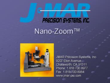
Nano-Zoom
- JMAR Precision Systems, Inc.
- 9207 Eton Avenue,
- Chatsworth, CA 91311
- Phone 1.818.700.8977
- Fax 1.818.700.8984
- www.jmar-psi.com
2
Nano-Zoom
- The Nano-Zoom combines a high quality optical
microscope and a state-of-the-art scanning probe
microscope (SPM) into a single system. - The Nano-Zooms SPM is capable of
- Atomic Force Microscopy
- Lateral Force Microscopy
- Magnetic Force Microscopy (optional)
- Electric Force Microscopy (optional)
- Use the optical microscope to find a feature on
your sample. - Press a button to move that feature precisely
under the SPM. - Scan your sample with the SPM at Angstrom
resolution. - The Nano-Zoom can be equipped with a vacuum
chuck for holding disk media or semiconductor
wafers, or fixtures for a variety of other
materials.
3
Nano-Zoom
4
Why use an AFM?
- Resolution.
- The resolving power of an optical microscope is
about 300 nm. - AFM offers the best resolution and fidelity. The
sharp, crisp high definition edge sets the stage
for good measurement. - Material independence.
- Versatility.
- Surface topography
- Height measurements
- Width measurements
- Profiles
- Non-Destructive.
- Compare to SEM, whose cross section measurements
are destructive.
5
How Does an AFM Work?
- Instead of using light or electrons to probe the
sample, the AFM uses a tip suspended above the
surface. - The attractions or repulsions between the tip and
the surface cause the tip to deflect. - A laser senses the deflection.
- Scanning the tip across the surface generates the
image.
6
Scanned Disk Media
- The next slide shows a 5 mm 5 mm Nano-Zoom
Atomic Force Microscope (AFM) image of rigid disk
media. The AFM renders a 3-dimensional image of
the surface of the media. Features that are too
small to see in an optical microscope are clearly
visible in the AFM scan.
7
AFM Image of Disk Media
8
AFM Image of Disk Media 3D View
The next slide shows a 3 dimensional plot of the
same 5 mm 5 mm AFM scan of rigid disk media.
9
AFM Image of Disk Media -- 3D View
10
Surface Roughness Calculation
- The Nano-Zoom includes software with many
features for analyzing AFM data. The next slide
shows a height histogram and surface roughness
calculations for the same AFM scan of rigid disk
media. For this sample, the RMS deviation of the
surface height is 4 Angstroms.
11
Surface Roughness CalculationAFM Data on Rigid
Disk Media
12
AFM Image of Grating Pattern
- The next slide shows an AFM image of a metallic
grating imprinted on a film substrate.
13
AFM Image of Grating Pattern Slope View
14
AFM Image of Grating Pattern 3D View
- The next slide shows a 3 dimensional rendering of
the same AFM scan.
15
AFM Image of Grating Pattern 3D View
16
Measurement View of Grating Pattern
- The next slide shows the measurement screen of
the AFM software, along with a sample measurement
of two periods of the grating. The grating pitch
is 40 mm, conforming to specification!
17
Measurement View of Grating Pattern
18
Defect View
- The next slide shows the Defect Screen of the
Nano-Zoom software. Several software features
are illustrated, including the following - A defect table with imported data.
- A Polar View of the entire sample surface
indicating the current field of view and the
defect locations. - Live video from the microscope.
- Big Digital Readouts showing the position in the
sample parts coordinate system.
19
Nano-Zoom Defect View
20
Nano-Zoom Applications
- Defect Review
- CMP Process Verification
- IC Failure Analysis
- Width, Depth, and Height Measurements
- Surface Roughness
- Fiber Optic Gratings
- Pole Tip Recession
- CD/DVD Inspection
- Disk Media Inspection
- Wafer Inspection
- Thin Film Inspection
- Etc.
21
Specifications AFM Optics
- Scanning Probe Microscope (SPM) Specifications
- X,Y Scan Size 80 µm x 80 µm standard, extendable
to 200 µm. - Z Range 8 µm standard, extendable to 20 µm.
- X,Y resolution 1 nm (smaller resolutions are
available). - Z resolution lt 1 Å.
- Variety of probe tips available
- Operational modes Contact, Intermittent Contact,
Broadband, etc. - Lateral Force Microscopy.
- Optional Magnetic Force Microscopy.
- Optional Electric Force Microscopy.
- Easy probe tip installation and alignment.
- Automatic probe tip engage feature.
- Top Quality Optical Microscope
- Dark field and bright field illumination.
- 5 position lens turret.
- Objective lenses 5x, 20x, 50x, 100x
standard.Other lenses are available. - 10X eyepieces standard.
- Optional Differential Interference Contrast
(DIC).
22
Additional Specifications
- Precision Motion Platform
- Computer-controlled R, ?, Z precision motion
platform. - R Stage 0.1 micron resolution, 10 inches travel.
- Z Stage 0.1 micron resolution, 6 inches travel.
- ? Stage 0.001 resolution.
- For 150 mm wafers or disks (extendible to 200
mm). - Acoustic and vibration isolation included.
- Other features
- Optional scriber with diamond or steel tips to
etch your sample. - Acoustic and vibration isolation.
- Small equipment foot print.
- Measurements are non-destructive.
23
System Isolation
- Vibration Isolation
- Standard passive vibration isolation system,
natural frequency 1 Hz. - Optional AVI-150 active vibration isolation
system. - Active isolation 1-200 Hz.
- Passive isolation gt200 Hz.
- Correctional forces to 8N horizontal, 4N
vertical. - Isolation in 6 degrees of freedom.
- Virtually no resonance.
- Acoustic Isolation
- Foam-lined enclosure isolates the system against
acoustic noise and air currents.
Optional AVI-150
24
Software Features
- Easy to Use Windows-based software.
- 3D Visualization of AFM images.
- Image refinement with tilt removal, streak and
spot removal, smoothing and user defined filter. - Fourier transform analysis for analyzing and
modifying the frequency spectrum of SPM images. - Histogram analysis and surface roughness
measurements. - Dimensional analysis for measuring feature
height, width, and length. - Image storage and retrieval.
- Optional Pax-It image archival software
maintains a database of images, makes it easier
to organize, analyze, network, transmit and print
images. - Defect classification system. Defect table
displays list of defects, allows user to move to
a defect manually or automatically and classify
it. - Reads/writes text files containing defect
positions. Easy to export/import defect data
to/from other machines. - Polar view displays a diagram of your sample with
all defect locations. - Large digital readouts display part position
continuously.
25
Choosing an AFM Probe Tip
Chose a probe tip with a small enough radius to
resolve features of interest.
Chose a probe tip that is long enough and with a
narrow enough cone angle for the features being
measured.
26
JMAR Probe Tips
27
JMAR-PSI Sales
- Address
- JMAR Precision Systems, Inc.
- 9207 Eton Ave.
- Chatsworth, CA 91311-5808
- Web
- www.jmar-psi.com
- salesjpsi_at_jmar-psi.com
- Phone
- 1-800-793-0179
- 1-818-700-8977
- Fax
- 1-818-700-8984
28
Image Gallery
The remaining slides have more SPM images and
measurements. Enjoy!
29
Alumina Layer
30
CeO2 Film
31
Crystals on Pr-Ce Pellet Surface
32
Sapphire Substrate
33
SiO2 Film
34
SiO2 on Re Crystal Substrate
35
Northrop-Grumman Test Pattern
36
Alumina Layer With Vias
37
Line Heights
38
Pits on CD Media
39
Via Height Measurements
40
Photoresist Residue on 1-D Refill Lithography
41
GaN
42
Homoepitaxial SiC
43
Polished SiC
44
SiC Film
45
Corrosion
46
Chrome on Glass
47
Stainless Steel
48
Ti Film
49
JMAR-PSI Sales
- Address
- JMAR Precision Systems, Inc.
- 9207 Eton Ave.
- Chatsworth, CA 91311-5808
- Web
- www.jmar-psi.com
- salesjpsi_at_jmar-psi.com
- Phone
- 1-800-793-0179
- 1-818-700-8977
- Fax
- 1-818-700-8984


