Developmental biology - PowerPoint PPT Presentation
1 / 48
Title:
Developmental biology
Description:
Draw out embryonic development from 1-cell stage, cleavage, blastula, gastrula, ... (urochordate (tunicate) (notochord in larvae), hydra (cnidaria, invertebrate) ... – PowerPoint PPT presentation
Number of Views:220
Avg rating:3.0/5.0
Title: Developmental biology
1
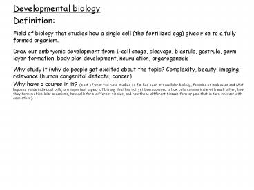
- Developmental biology
- Definition
- Field of biology that studies how a single cell
(the fertilized egg) gives rise to a fully formed
organism. - Draw out embryonic development from 1-cell stage,
cleavage, blastula, gastrula, germ layer
formation, body plan development, neurulation,
organogenesis - Why study it (why do people get excited about the
topic? Complexity, beauty, imaging, relevance
(human congenital defects, cancer) - Why have a course in it? (most of what you have
studied so far has been intracellular biology,
focusing on molecules and what happens inside
individual cells one important aspect of biology
that has not yet been covered is how cells
communicate with each other, how they form
multicellular organisms, how cells form different
tissues, and how these different tissues form
organs that in turn interact with each other).
2
- Questions
- axis determination (AP, DV, LR) Q. how do you
break the symmetry of the egg? Sperm entry,
localized determinants - cell differentiation, cell proliferation, cell
growth - cell migration, polarization (symmetric vs
asymmetric cell division), cell shape (giving
cells different morphologies, e.g. neurons, EMT),
cell death - morphogenesis (how cells come together to form
tissues and how these tissues migrate) - organogenesis
- timing (biological clock)
- aging
- germ cell development, fertilization
- stem cells (what are they (multipotency, ability
to self-renew), why did they become so trendy
(Dolly the sheep, 1997, showed that cloning was
possible development of human ES cells, 1998
nobel for mouse ES cells induced pluripotent
cells )) - regeneration
- evo-devo (a field of biology that compares the
developmental processes of different animals in
an attempt to determine the ancestral
relationship between organisms and how
developmental processes evolved.) - Q. How efficient is human development? (talk
about implantation)
3
Human reproduction is an inefficient process
50 of concepti do not implant (implantation 8-10
dpf, Heart beat at 21 dpf). a further 30 die
and abort after implantation.
3-4 of all live births possess a
macroscopically visible congenital defect
(120,000 babies/year in the USA).
1 of all babies are born with a heart defect.
20 of neonatal deaths are caused by congenital
defects (the leading cause of neonatal death in
the USA)
congenital disorders are the cause of 50 of
pediatric admissions in the USA
Developmental defects seen at birth are caused by
defects in the cellular processes of development.
4
- Developmental biology
- Model systems
- Worm (C. elegans)
- Fly (Drosophila melanogaster)
- Fish (zebrafish, medaka)
- Mouse
- Xenopus (laevis, tropicalis)
- Chick (Quail)
- Sea urchin (echinoderm), ascidians (ciona
intestinalis, savigni) (urochordate (tunicate)
(notochord in larvae), hydra (cnidaria,
invertebrate), planarians (flatworm), arabidopsis
(weed) ( other plants), axolotls (and other
salamanders), chlamydomonas (unicellular green
alga, protozoan), yeast (?), - Many systems available which one to use most
powerful one where you can study your questions
of interest. (also, many of these model systems
are used to study processes other than
developmental biology).
5
Friday, May 30th 17th Annual Developmental
Biology Symposium Developmental insights
from atypical model systems Speakers
Eduardo R. Macagno, Ph.D., University of
California, San Diego (leech) Gary Wessel,
Ph.D., Brown University (starfish) William
Smith, Ph.D., University of California, Santa
Barbara (ascidians) Richard Behringer, Ph.D.,
University of Texas (Bats) Kathleen K. Smith,
Ph.D., Duke University (marsupials) Host
The Developmental Biology Journal Club Rock
Hall Auditorium
6
- Developmental biology
- Tools
- - Forward genetics
- - Reverse genetics (is gene x necessary?)
(mutation, RNAi, antisense oligos) - - Gain-of-function expts (is gene x sufficient)
(mRNA injections, transgenics various levels of
sophistication, gal4-uas, temporal and spatial
control) - - Where is function of gene x required? Creating
genetic mosaics (genetic, transplants) - Imaging (live, gene and protein expression)
- - Experimental embryology (cell or tissue
ablations, bead implantation, chick-quail
chimeras)
7
(No Transcript)
8
C. elegans (adult 1 mm in length)
9
C. elegans development (embryo about 50 ?m in
length)
10
Xenopus laevis
AP
V
D
VP
D
A
P
V
11
Early developmental stages of Xenopus laevis
2.5 hpf
5 hpf
10 hpf
3.5 hpf
hpf hours post-fertilization
12
Early developmental stages of Xenopus laevis
Cleavage movie
13
Developmental stages of Xenopus laevis
14
Gastrulation the ultimate cell migration problem
15
Gastrulation and Neurulation highly
coordinated tissue movements
Keller neurulation movie
16
(No Transcript)
17
Zebrafish embryonic development
18
http//anatomy.ucsf.edu/devbio/course.htm
19
(No Transcript)
20
http//www.nature.com/milestones/development/miles
tones/index.html
21
The Question
1 cell type
200 different cell types
22
Answer by the blastula stage (2000 cells)
- Evidence
- morphological differences
- fate map data
- e.g. ventral cells give rise to blood
- dorsal cells give rise to notochord
- - gene expression
23
Answer at the blastula stage (2000 cells)
- Evidence
- morphological differences
- fate map data
- e.g. ventral cells give rise to blood
- dorsal cells give rise to notochord
- - gene expression
AP animal pole VP vegetal pole D dorsal V
ventral
24
The turning point of Hans Spemanns career
partial constriction
25
Hilde Mangold and Hans Spemann, 1924
26
Hilde Mangold and Hans Spemann, 1924
a
27
(No Transcript)
28
(No Transcript)
29
Tissue from the dorsal side of the embryo
(dorsal lip of the blastopore) is transplanted
to the ventral side of the embryo.
This induces cells on the ventral side of the
embryo to form a second neuraxis ventrally
30
Modern version of the Mangold and Spemann
experiment
Gastromaster movie http//www.xenbase.org/methods/
methods.html
31
What is wrong with this representation of the
Mangold and Spemann experiment?
32
(No Transcript)
33
A small region of the embryo, the organizer or
dorsal lip, has the ability when grafted to the
opposite (ventral) side to cause pronounced
cell non-autonomous effects. Neighboring cells
in the ectoderm are induced to form central
nervous system and those in the mesoderm to form
dorsal structures such as somites. These
inductive effects should be mediated by
diffusible molecules and the isolation of the
molecular signals involved has been a challenge
for generation of embryologists (Viktor
Hamburger, 1988).
Seeking the molecules responsible for the
activity of the Organizer (using the tools
available in late 1980s). a
34
Seeking the molecules responsible for the
activity of the Organizer I. Inducing the
formation of an ectopic axis
Inject RNA (from Organizer) into
ventrovegetal blastomere
V
D
Lateral view
Formation of an ectopic axis
Dorsal view
35
- Richard Harland (UC Berkeley)
- used an expression screen for activities that
induce - dorsal/head structures in Xenopus embryos
- - identified Noggin (Smith and Harland, Cell,
1992)
- Eddy De Robertis (UCLA)
- used an expression screen for activities that
rescue - UV ventralized Xenopus embryos
- - identified Chordin (Sasai et al., Cell, 1994)
Two novel secreted proteins. What next?
36
Data suggesting that Noggin and Chordin could
function by antagonizing BMP signaling.
BMPs act as ventralizers and are expressed in
the ventral side of the embryo (1992)
DN-BMP receptors have dorsalizing activity (1994)
The Drosophila homologue of Chordin
(Short-gastrulation) functions genetically as an
antagonist of Dpp (BMP homologue)
37
The model
38
Additional members of the pathway were initially
identified genetically in Drosophila
Tolloid (clearly functions positively in BMP
signaling)
39
Xolloid (Xenopus tolloid) cleaves Chordin,
reducing its affinity for BMP, thereby allowing
BMP binding to its receptor
40
Model of BMP4 gradient leading to different
mesodermal cell types
41
What is responsible for the formation of
the dorsal organizer?
42
Embryologically By 4 cell stage, the embryo is
asymmetric in its potential (Spemann, 1938)
43
Embryologically By 4 cell stage, the embryo is
asymmetric in its potential (Spemann, 1938)
44
- Molecularly
- Wnt pathway implicated
- Wnt injection leads to secondary axis
- (Andy McMahon and Randy Moon, Cell, 1989)
- depletion of beta-catenin inhibits dorsal tissue
formation - (Janet Heasman, Chris Wylie, et al. Cell, 1994)
- Wnt11 appears to be the initiating event
- (Janet Heasman, Chris Wylie, et al. Cell, 2005)
45
Wnt signaling, 2006 Xenopus assays, fly
genetics, yeast 2-hybrid
http//www.stanford.edu/rnusse/wntwindow.html
46
http//faculty.washington.edu/rtmoon/cell2.html
47
Proteins secreted by the dorsal and ventral
signaling centers in the Xenopus gastrula
48
- Summary
- Key concepts
- Induction (organizing centers)
- Conservation of signaling pathways (vertebrates,
fly)
- Activators and Inhibitors (negative feedback
loops)
- Complexity


![[PDF] DOWNLOAD EBOOK Human Embryology and Developmental Biology PowerPoint PPT Presentation](https://s3.amazonaws.com/images.powershow.com/10130801.th0.jpg?_=20240916076)


![[PDF] DOWNLOAD FREE Human Embryology and Developmental Biology - Inkli PowerPoint PPT Presentation](https://s3.amazonaws.com/images.powershow.com/10130802.th0.jpg?_=20240916075)


