Virtual Radiographic Environments - PowerPoint PPT Presentation
1 / 1
Title:
Virtual Radiographic Environments
Description:
The goal is to create a software tool for computer assisted instruction ... computed tomography (CT) data, Forensic Science International, 71, 199-204 ... – PowerPoint PPT presentation
Number of Views:28
Avg rating:3.0/5.0
Title: Virtual Radiographic Environments
1
Virtual Radiographic Environments Philip Cosson,
Guillaume Debouzy, Daniel Deprez, Franck Vidal,
Suresh Keswani, Julian Warren. School of Health
and Social Care and School of Computing and
Mathematics University of Teesside
Abstract
Computer software was created to simulate the
process of administering an x-ray, from the
initial positioning of patient, x-ray source and
cassette, to the resulting image. Thanks to this
approach patient exposure is no longer necessary
for training and research. Two programs have
already been created one for research and
another for teaching purposes.
Introduction The goal is to create a software
tool for computer assisted instruction realistic
enough to be used in radiography education and
research. The current method of training
individuals to work in any clinical environment
requires some hands-on experience1. Training
workers to handle radiation sources is
particularly challenging because the danger is
not always visually apparent2. Existing software3
has been developed for scientific rather than
educational use.
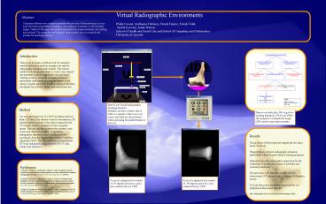
Here is our Virtual Radiographic Teaching
Interface. Students can have a better idea of
what is a cassette, what is an x-ray source and
what are the problems with positioning the
patient thanks to this tool.
Method Our software makes use of a DICOM dataset
derived from a CT scan. The dataset is used to
reconstruct a 3D surface rendered model of the
object scanned by the CT scanner, which is
displayed on the computer screen. The user can
then position the cassette, x-ray source and
object as required. A simulated radiographic
exposure control panel passes the coordinates
from the interface to a Simple Projection
Algorithm (SPA). The SPA calculates a
conventional 2D X-ray summation image from the 3D
CT data volume and displays it.
Here is our work plan. Weve got two working
interfaces (VRTI and VRRI). The program to
compute the image (SPA) needs some improvements.
- Results
- The products of this project are targeted at two
main areas of activity - Supporting pre-clinical radiography education,
particularly within Enquiry Based Learning
programs. - Research into new radiographic projections for
the evaluation of disease and trauma in medicine
and veterinary medicine. - The next step a 3D interface, usable with 3D
stereoscopic LCD glasses and a 6-degrees-of-freedo
m mouse. - You can learn more about this project and try our
programs at the project website - http//radiography.tees.ac.uk/soh_research/html/pa
ge-1.html
References 1) Riepert T Ulmcke D Jendrysiak U
Rittner C (1995) Computer-assisted simulation of
conventional roentgenograms from
three-dimentional computed tomography (CT) data,
Forensic Science International, 71, 199-204 2)
Michel MS Knoll T Kohrmann KU Alken P (2002)
Development and evaluation of a new
computer-based interactive training system for
virtual life-like simulation of diagnostic and
therapeutic endourological procedures. BJU
International. 89(3)174-7, 2002 3) Hahn JK
Kaufman R Winick AB Carleton T. Park Y Lindeman
R Oh KM (1998) Training environment for inferior
vena caval filter placement Studies in Health
Technology Informatics. 50291-7, 1998.
X-ray of a phantom foot rotated of 90 degrees
about its y axis created with our VRTI
X-ray of a phantom foot rotated of -90 degrees
about its x and y axis created with our VRTI


