Functional Magnetic Resonance Imaging fMRI - PowerPoint PPT Presentation
1 / 25
Title:
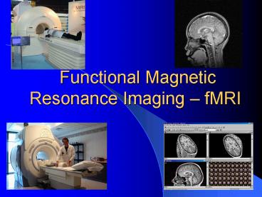
Functional Magnetic Resonance Imaging fMRI
Description:
... observed in the United States in 1946 by Felix Bloch, William W. Hansen, and ... Felix Bloch and Edward Purcell shared the 1952 Nobel Prize in physics for it. ... – PowerPoint PPT presentation
Number of Views:149
Avg rating:3.0/5.0
Title: Functional Magnetic Resonance Imaging fMRI
1
Functional Magnetic Resonance Imaging fMRI
2
Magnetic Resonance
- Magnetic Resonance is the absorption or emission
of electromagnetic radiation by electrons or
atomic nuclei in response to the application of
magnetic fields. - Magnetic Resonance is divided into Electron
Magnetic Resonance or EMR where electrons absorb
or emit electromagnetic radiation and into
Nuclear Magnetic Resonance or NMR where nuclei
(or protons) absorb or emit electromagnetic
radiation
3
- Nuclear Magnetic Resonance (NMR) of protons was
first observed in the United States in 1946 by
Felix Bloch, William W. Hansen, and Martin E.
Packard at Stanford University and independently
by Edward M. Purcell, Robert V. Pound, and Henry
C. Torrey at Harvard. - Felix Bloch and Edward Purcell shared the 1952
Nobel Prize in physics for it. NMR quickly proved
an invaluable tool for identifying chemical
compounds and for studying their molecular
structures. - NMR also forms the basis for MRI, which is used
in medicine since the 1980s.
Felix Bloch
Edward Purcell
4
NMR involves three fundamental physical phenomena
related to magnetic fields.
5
First Phenomenon
- The movement of charged particles gives rise to a
magnetic field. - A nucleus behaves like the spinning (moving) ball
of positive charge it will therefore create a
magnetic field. - The direction of a nucleuss magnetic field is
closely associated with that of its spin axis
curl your right hand around the nucleus of a
hydrogen atom, say, in the direction in which it
is rotating then your thumb defines the
direction of the spin, and points out of the
protons north magnetic pole.
6
Second Phenomenon
- A magnetized body placed in a magnetic field
already in existence will experience a torque, or
twisting force, and try to align along that
field.
7
Third Phenomenon
- A nucleus has the option to be in a quasi-stable,
high-energy state, spinning in the wrong
direction, with its nuclear magnetic field
pointing anti-parallel to the external field. - The stronger the magnetic field of the magnetized
body, the harder it will be to twist it over so
that it points in the wrong direction (i.e.
anti-parallel to the external field) the
greater the effort that is required. - Likewise, the energy needed to flip over the
magnetized body will increase with the strength
of the external magnetic field.
8
- One process by which a nucleus can be elevated
from the lower- to the higher- energy spin state
(be flipped over such that it is anti-parallel to
the external field) is through the absorption of
a photon of the right energy - The energy difference between the high
(anti-parallel to the external field) and low
(parallel to the external field) energy protons
is measurable and is expressed in the Larmor
equation - Resonance is referred to as the property of an
atom to absorb energy only at the Larmor
frequency.
9
- In NMR the resonance (the energy absorbs or
emitted to move nuclei from low to high state of
energy and vise versa, respectively) of
different material in different environments is
monitored in order to analyze the molecular
composition of the material in question.
10
Relaxation Times
- The time it takes a nucleus to emit the energy it
absorbed and to return to its low energy state is
termed the relaxation time. - Relaxation time is another, very important,
measure in NMR and in MRI.
11
Magnetic Resonance Imaging - MRI
- Raymond Damadian a physician at Downstate Medical
Center in Brooklyn, New York, found in 1971 that
tissues surgically removed from different organs
in rats have significantly different relaxation
times and that tumors in some organs tend to have
measurably longer NMR relaxation times than do
the corresponding healthy tissues - That was the first step toward the medical use of
NMR
12
- Soon thereafter Damadian and Paul Lauterbur
separately suggested ways in which the NMR
signals coming from different parts of the body
could be untangled from one another and utilized
in imaging. - Damadian completed the first whole-body MRI
scanner (named indomitable, now on display at
the Smithsonian Museum) in 1977. - These early efforts, followed by the work of
hundreds of others, have led to the development
of MRI machines that can now map out spatial
variations in relaxation times in great detail
and with superb soft-tissue contrast. MRI can
thus provide useful information not just about
anatomy, but also about the physiology of cells,
and even about their health.
13
- MRI can produce three-dimensional anatomical
images of thin tissue slices at any orientation.
- MRI can reveal valuable information on the
metabolic and physiological status of soft
tissues. - In MRI there are no radiation risks to the
patient there are no X rays, MRI involves only
stable, non-radioactive nuclei and a variety of
magnetic fields.
14
(No Transcript)
15
- Approximately 70 of the body is made up of water
which contains two hydrogen atoms and one oxygen
atom. - The nucleus of the hydrogen atom consists of one
proton, and no neutrons. - The single proton in the nucleus of the hydrogen
atom gives it a positive charge and creates a
large magnetic moment (i.e. magnetic field). - All the above make hydrogen atoms extremely
sensitive to magnetic resonance. - Indeed, it is the hydrogen atoms that are focused
on to produce a MR image.
16
Precession
- Hydrogen atoms do not actually align directly
with the direction of the magnetic field, but
rather rotate or wobble around the axis of the
external magnetic field (B0). What is actually
aligned with the axis of B0 is the axis around
which the hydrogen proton wobbles. - The term to describe this secondary spin is
precession. Protons actually precess at an angle,
spinning a cone-shape around the direction of the
external magnetic field. The speed at which the
protons precess is referred to as the
precessional frequency and is measured in
megahertz.
Spinning Proton
Precession
17
MRI utilize three different magnetic fields
- The principal or external field
- The gradient magnetic fields
- The magnetic component of radio frequency fields
18
The Principal or External Field
- The external field generated by the principal
magnet, is designed to be very strong ,
homogenous (uniform) throughout the volume of the
patients body being imaged, and constant over
time. - Magnetic field strength is measured in a unit
called the Tesla (T). The field strength of the
earth itself, viewed as a huge bar magnet, is
about 0.00005 T. - MRI is commonly carried out with the principal
field in the 0.1 T to 1.5 T range.
19
The Gradient Magnetic Fields
- The gradient magnetic fields, generated by the
gradient magnets, are intentionally made to vary
in strength from place to place, and are rapidly
switched on and off intermittently from time to
time. - This allows a better localization of the signal
in MRI
20
The Magnetic Component of Radio Frequency Fields
- The magnetic component of radio frequency fields,
generated by the radio wave transmission and
reception equipment, oscillate millions of times
a second. - In MRI the radio wave transmission equipment
broadcasts a radiofrequency (RF) pulse toward the
human body. This pulse frequency is the same as
precess frequency of the hydrogen protons (i.e.
at the hydrogen Larmor frequency) and
consequently it elevates them to a higher energy
state. In other words, the hydrogen protons
"resonate" to the pulse frequency.
21
T1 T2 Relaxation Times
- T1 is the time it takes the orientation of the
spin to be again parallel to the magnetic field
created by the large magnet. - T2 is the time it takes the radius of the spin
itself to become less wobbly and like the
original spin.
22
- The values of T1 and T2 are strongly influenced
by the precise manner in which the water
molecules are moving around and bumping into
other molecules within the cells. - Those interactions, in turn, are highly sensitive
to the fine-tuning of the physiology of the
tissues. - T1 and T2 have differential sensitivity to
different tissues. - Therefore, MRI can produce images that clearly
distinguish among the various parts of brain
tissue and other organs.
23
fMRI
- In 1990, Seiji Ogawa of ATT's Bell Laboratories
reported that, in studies with animals,
deoxygenated hemoglobin, when placed in a
magnetic field, would increase the strength of
the field in its vicinity, while oxygenated
hemoglobin would not. - Ogawa showed in animal studies that a region
containing a lot of deoxygenated hemoglobin will
slightly distort the magnetic field surrounding
the blood vessel, a distortion that shows up in a
magnetic resonance image.
24
- Functional MRI is based on the increase in blood
flow to the local blood vessels that accompanies
neural activity in the brain. - This results in a corresponding local reduction
in deoxyhemoglobin because the increase in blood
flow occurs without an increase of similar
magnitude in oxygen extraction - Deoxyhemoglobin alters the T2 weighted magnetic
resonance image signal. - Thus, deoxyhemoglobin is sometimes referred to as
an endogenous contrast enhancing agent, and
serves as the source of the signal for fMRI.
25
(No Transcript)


![[PDF] Handbook of Functional MRI Data Analysis 1st Edition, Kindle Edition Kindle PowerPoint PPT Presentation](https://s3.amazonaws.com/images.powershow.com/10077861.th0.jpg?_=20240712083)