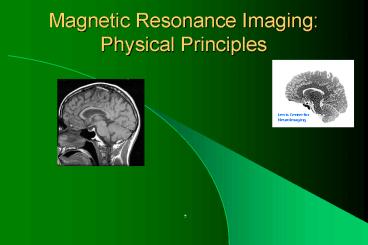
Magnetic Resonance Imaging: Physical Principles - PowerPoint PPT Presentation
1 / 58
Title:
Magnetic Resonance Imaging: Physical Principles
Description:
Felix Bloch & Edward Purcell NM resonance. Ernst & Anderson FT NMR ... Bloch, Purcell and Ernst have been awarded the Nobel Prize for their work. 7/8/09. 3 ... – PowerPoint PPT presentation
Number of Views:341
Avg rating:3.0/5.0
Title: Magnetic Resonance Imaging: Physical Principles
1
Magnetic Resonance ImagingPhysical Principles
Lewis Center for NeuroImaging
- ,
2
A very brief history
- Stern and Gerlach atomic beam experiments
- Isidor Rabi molecular beam exp. nuclear
magnetic moments (angular momentum) - Felix Bloch Edward Purcell NM resonance
- Ernst Anderson FT NMR
- Ernst, Aue, Jeener, et al 2D FT NMR
- Lauterbur Mansfield NMR imaging
- Bloch, Purcell and Ernst have been awarded the
Nobel Prize for their work
3
Physics of MRI, An Overview
- Nuclear Magnetic Resonance
- Nuclear spins
- Spin precession and the Larmor equation
- Static B0
- RF excitation
- RF detection
- Spatial Encoding
- Slice selective excitation
- Frequency encoding
- Phase encoding
- Image reconstruction
- Fourier Transforms
- Continuous Fourier Transform
- Discrete Fourier Transform
- Fourier properties
- k-space representation in MRI
4
Physics of MRI
- Echo formation
- Vector summation
- Phase dispersion
- Phase refocus
- 2D Pulse Sequences
- Spin echo
- Gradient echo
- Echo-Planar Imaging
- Medical Applications
- Contrast in MRI
- Bloch equation
- Tissue properties
- T1 weighted imaging
- T2 weighted imaging
- Spin density imaging
- Examples
5
Larmor Revisited
6
Nuclear Precession and the Larmor Relationship
7
Alignment in an Applied Magnetic Field
Bo
8
NMR/MRI Sensitivity
- E h ? B
- N-/N e-E/kT 7 x10⁻⁶ at 1T for protons
The higher the field, the larger the net
magnetization and the bigger the MR signal !!!!
9
Many spins in a voxel vector summation
spins not in step
spins in step
Rotating frame Lamor precession
10
Phase dispersion due to perturbing B fields
Spin Phase f ? gBt B B0 dB0 dBcs dBpp
sampling
sometime after RF excitation
Immediately after RF excitation
11
Refocus spin phase echo formation
time
Echo Time (TE)
- Invert perturbing field dB -dB
- Invert spin state f -f
Phase 0 dBt f-dB(t-TE/2) 0
(gradient echo, k-space sampling)
Phase 0 dBt -fdB(t-TE/2)
0
(spin echo)
12
Spin Echo
- Spins dephase with time
- Rephase spins with a 180 pulse
- Echo time, TE
- Repeat time, TR
- (Running analogy)
13
Frequency encoding - 1D imaging
Spatial-varying resonance frequency during RF
detection
B B0 Gxx
- S(t) eigBt
- S(t) ?m(x)eigGxxtdx
m(x)
kx gGxt
x
S(t) ?m(x)eikxxdx S(kx), m(x) FTS(kx)
14
Slice selection
Spatial-varying resonance frequency during RF
excitation
w w0 gGzz
w
B1 freq band
z
Excited location
Slice profile
m mximy g ?b1(t)e-igGzztdt B1(gGzz)
15
Gradient Echo FT imaging
ky
Readout
kx
Repeat with different phase-encoding amplitudes
to fill k-space
16
EPI (echo planar imaging)
X
ky
Y
Z
kx
RF
time
Quick, but very susceptible to artifacts,
particularly B0 field inhomogeneity. Can acquire
a whole image with one RF pulse single shot EPI
17
Spin Echo FT imaging
ky
Readout
kx
Repeat with different phase-encoding amplitudes
to fill k-space
18
Spin Relaxation
- Spins do not continue to precess forever
- Longitudinal magnetization returns to equilibrium
due to spin-lattice interactions T1 decay - Transverse magnetization is reduced due to both
spin-lattice energy loss and local, random, spin
dephasing T2 decay - Additional dephasing is introduced by magnetic
field inhomogeneities within a voxel T2' decay.
This can be reversible, unlike T2 decay
19
Bloch Equation
- The equation of MR physics
- Summarizes the interaction of a nuclear spin with
the external magnetic field B and its local
environment (relaxation effects)
20
Contrast - T1 Decay
- Longitudinal relaxation due to spin-lattice
interaction - Mz grows back towards its equilibrium value, M0
- For short TR, equilibrium moment is reduced
21
Contrast - T2 Decay
- Transverse relaxation due to spin dephasing
- T2 irreversible dephasing
- T2/ reversible dephasing
- Combined effect
22
Free Induction Decay Gradient echo (GRE)
- Excite spins, then measure decay
- Problems
- Rapid signal decay
- Acquisition must be disabled during RF
- Dont get central echo data
MR signal
e-t/T2
time
0
90 RF
23
Spin echo (SE)
e-t/T2
MR signal
e-t/T2
time
24
MR Parameters TE and TR
- Echo time, TE is the time from the RF excitation
to the center of the echo being received.
Shorter echo times allow less T2 signal decay - Repetition time, TR is the time between one
acquisition and the next. Short TR values do not
allow the spins to recover their longitudinal
magnetization, so the net magnetization available
is reduced, depending on the value of T1 - Short TE and long TR give strong signals
25
Contrast, Imaging Parameters
26
Properties of Body Tissues
MRI has high contrast for different tissue types!
27
MRI of the Brain - Sagittal
T1 Contrast TE 14 ms TR 400 ms
T2 Contrast TE 100 ms TR 1500 ms
Proton Density TE 14 ms TR 1500 ms
28
MRI of the Brain - Axial
T1 Contrast TE 14 ms TR 400 ms
T2 Contrast TE 100 ms TR 1500 ms
Proton Density TE 14 ms TR 1500 ms
29
Brain - Sagittal Multislice T1
30
Brain - Axial Multislice T1
31
Brain Tumor
T1
T2
Post-Gd T1
32
2D Sequence (Gradient Echo)
ky
acq
Gx
Gy
kx
Gz
b1
TE
Scan time NyTR
TR
33
Spectroscopy
- Precession frequency depends on the chemical
environment (dBcs) e.g. Hydrogen in water and
hydrogen in fat have a ?f fwater ffat 220
Hz - Single voxel spectroscopy excites a small (cm3)
volume and measures signal as f(t). Different
frequencies (chemicals) can be separated using
Fourier transforms - Concentrations of chemicals other than water and
fat tend to be very low, so signal strength is a
problem - Creatine, lactate and NAA are useful indicators
of tumor types
34
BOLD functional MRI
Magnetic properties of oxyhemoglobin and
deoxyhemoglobin L. Pauling and C. Coryell, PNAS
USA 22210-216 (1936) BOLD effects in vivo S.
Ogawa, et al., MRM, 1468-78 (1990) BOLD
activation experiments K. K. Kwong, et al., PNAS
USA, 895675-5679 (1992) S. Ogawa, et al., PNAS
USA, 895951-5955 (1992) P. A. Bandettini, et
al., MRM, 25390-397 (1992) J. Frahm, et al.,
JMRI, 2501-505 (1992)
35
Mechanism of BOLD Functional MRI
Brain activity
Oxygen consumption
Cerebral blood flow
Oxyhemoglobin Deoxyhemoglobin
Magnetic susceptibility
T2
MRI signal intesity
36
Magnetic Properties of Oxyhemoglobin and
Deoxyhemoglobin
Deoxyhemoglobin paramagnetic (c gt 0)
paramagnetic with respect to the surrounding
tissue Oxyhemoglobin diamagnetic (c lt 0)
isomagnetic with respect to the surrounding
tissue
37
Magnetic Susceptibility
38
Oxyhemoglobin and Deoxyhemoglobin in Veins during
Brain Activation
Activation
Rest
Normal blood flow
High blood flow
Oxyhemoglobin Deoxyhemoglobin
39
T2 Effect in fMRI
action
MR signal (S)
rest
TE
t
reception
excitation
40
Time Series and Activation Maps
Signal Intensity
Off
Off
Off
Off
On
On
On
On
Scan Number
41
Challenges in Functional FMRI
Sensitivity (Contrast-to-noise ratio) BOLD
signal change is 1-2 at 1.5 T signal-to-noise
ratio in single-shot EPI images is
100. Physiological pulsations (cardiac and
respiratory) Head motion instrumental
instability Specificity Location of activation
neurons or veins Susceptibility artifacts
42
Challenges in Functional FMRI
Temporal resolution Limited by BOLD
impulse-response function, image sampling rate,
and spin relaxation times Spatial
resolution Limited by BOLD point-spread
function, signal-to-noise ratio, and image
sampling rate Non-linearity Neurological and
hemodynamic Acoustic noise
43
Contrast-to-Noise Ratio
Brain Activation-related signal
change Sensitivity Temporal fluctuation
of image intensity
44
Enhancement of BOLD Contrast
Higher magnetic fields BOLD signal change DS Ba
(1 lt a lt 2) Standard clinical MRI scanner at 1.5
T Research scanner up to 8 T currently Optimizat
ion of image acquisition parameters Optimal echo
time (TE) to maximize BOLD signal Optimal
repetition time (TR) to increase number of images
acquired per unit time, and to decrease motion
artifacts
45
TE Dependence of Signal Change
TE
46
Suppression of Temporal Fluctuations
Head motion reduction Head holder modified from a
football helmet Image realignment in data
processing Physiological pulsations Correction
using simultaneously recorded cardiac and
respiratory signals Ultra-fast imaging
techniques Single-shot echo-planner imaging
(EPI) Single-shot Spiral imaging Post-processing D
enoising
47
Ultra-Fast Spiral Scanning
Fast An image (64x64) can be acquired in 20
ms Reduce head motion Increase number of images
collected per unit time Stable Spiral trajectory
is insensitive to motion and flow artifacts Zero
gradient moments at the center of k-space
(self-navigated) First-order gradient moments
vary smoothly over k-space
48
Multi-Slice Spiral Images
49
Activation Maps on Anatomical Images
MS Spiral
MS EPI
3D Spiral
50
Comparison of Activation Studies Using MS-spiral,
MS-EPI, and 3D-spiral
51
Temporal resolution
Impulse-response function
12s
5s
2s
t
52
Temporal resolution
- Sampling rate (single-shot EPI 10-15 slices/sec)
- Whole brain (30 4mm-slices) 2-3 sec
- T1 relaxation times
- Grey matter 1 sec
- White matter 0.8 sec
- CSF 2-3 sec
53
Spatial resolution
- BOLD point-spread function
- Spatial extent of neuronal activity, CBF, and
BOLD - Image spatial resolution
- 64x64 with FOV 240 mm 3.75mm
- 128x128 with FOV 240 mm 1.875 mm
- Signal-to-noise ration
- Single-shot EPI with voxel size 4x4x4 mm3 100
54
Non-linearity of BOLD Response
BOLD response vs. length of stimulation
t
2t
BOLD response during rapidly-repeated stimulation
ts
55
Experimental Designs in fMRI
Block-Design fMRI
Task
Rest
20-60s
Event-Related fMRI
8-12s
56
Hemodynamic Response vs. ISI
57
Visual Activation Maps (ISI12s)
58
Data Analysis Methods For fMRI
Hypothesis-driven approaches t-test,
cross-correlation, GLM, etc. Data-driven
approaches Principal component analysis (PCA),
independent component analysis (ICA), and
clustering analysis.


