Ultrasound Case: Shoulder glenohumeral joint Injection - PowerPoint PPT Presentation
Ultrasound Case: Shoulder glenohumeral joint Injection
Findings: Longitudinal extended field of view image of the quadriceps tendon ... The metal component (fc) of the femur (thin arrow) impinges the deep surface of ... – PowerPoint PPT presentation
Title: Ultrasound Case: Shoulder glenohumeral joint Injection
1
Ultrasound Case Shoulder (glenohumeral joint)
Injection
- Diagnosis Shoulder (glenohumeral joint)
injection - History Sonographic evaluation of a patient with
a total knee replacement - The presence of hardware does not interfere with
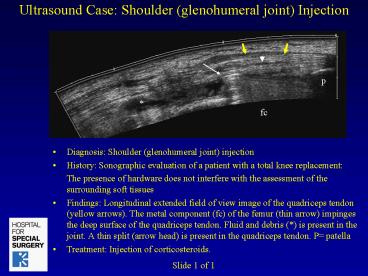
the assessment of the surrounding soft tissues - Findings Longitudinal extended field of view
image of the quadriceps tendon (yellow arrows).
The metal component (fc) of the femur (thin
arrow) impinges the deep surface of the
quadriceps tendon. Fluid and debris () is
present in the joint. A thin split (arrow head)
is present in the quadriceps tendon. P patella - Treatment Injection of corticosteroids.
Slide 1 of 1
PowerShow.com is a leading presentation sharing website. It has millions of presentations already uploaded and available with 1,000s more being uploaded by its users every day. Whatever your area of interest, here you’ll be able to find and view presentations you’ll love and possibly download. And, best of all, it is completely free and easy to use.
You might even have a presentation you’d like to share with others. If so, just upload it to PowerShow.com. We’ll convert it to an HTML5 slideshow that includes all the media types you’ve already added: audio, video, music, pictures, animations and transition effects. Then you can share it with your target audience as well as PowerShow.com’s millions of monthly visitors. And, again, it’s all free.
About the Developers
PowerShow.com is brought to you by CrystalGraphics, the award-winning developer and market-leading publisher of rich-media enhancement products for presentations. Our product offerings include millions of PowerPoint templates, diagrams, animated 3D characters and more.


