
Autophagy - PowerPoint PPT Presentation
1 / 26
Title:
Autophagy
Description:
3 After 4 days of treatment no more autophagosomes, only one large punctus ... Removal of K11777 (wash out) resulted in disappearance of autolysosome after 15 ... – PowerPoint PPT presentation
Number of Views:1923
Avg rating:3.0/5.0
Title: Autophagy
1
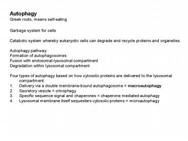
- Autophagy
- Greek roots, means self-eating
- Garbage system for cells
- Catabolic system whereby eukaryotic cells can
degrade and recycle proteins and organelles. - Autophagy pathway
- Formation of autophagosomes
- Fusion with endosomal-lysosomal compartment
- Degradation within lysosomal compartment
- Four types of autophagy based on how cytosolic
proteins are delivered to the lysosomal
compartment - Delivery via a double membrane-bound
autophagosome macroautophagy - Secretory vesicle crinophagy
- Specific sequence signal and chaperones
chaperone mediated autophagy - Lysosomal membrane itself sequesters cytosolic
proteins microautophagy
2
- Autophagy in more detail
- First described as a cellular response to
nutrient starvation (1992) - Four basic steps
- signaling
- sequestration of cytoplasm and vesicle formation
- docking and fusion of completed vesicle to
endosomal/lysosomal compartment - breakdown
- Understanding the molecular mechanism has been
revolutionized by whole genome sequencing and
subsequent analysis of gene deletion mutants,
especially in S. cerevisiae. - Autophagy related genes ATG
- Protein conjugation systems
- Most studies done in yeast and mammalian cells,
but other eukaryotic systems now studied too.
Importance of autophagy for host-pathogen
interactions is emerging (destruction of viruses
and bacteria).
3
Endosome sorting and autophagy are essential for
differentiation and virulence of Leishmania
major Besteiro, Williams, Morrison, Coombs,
Mottran JBC April 21, 2006 Analyzed expression
of L.major vacuolar sorting protein VSP4 by
expressing dominant mutant defective in
endosomal sorting and differentiation Localization
of GFP-ATG8 ATG4 knock-out (ATG4 is a cysteine
protease that processes ATG8) defective in
processing autophagosomes, vulnerable to
starvation, failed to undergo metacyclogenesis
4
(No Transcript)
5
1.Identification of candidate autophagy genes in
Leishmania major
Searched L.major genome with S. cerevisiae ATG
proteins (ATG genes autophagy related Arose from
whole genome sequencing and subsequent analysis
of gene deletion mutants)
Componenents of SNARE machinery (involved in
fusion of autophagosomes with vacuoles (yeast) or
endosomes (mammalian cells)) were also found
Overall conclusion from in silico
analysis L.major contains proteins involved in
autophagy, similarities to yeast and mammalian
cells. But some sequences have low similarities
(other pathways?) May be more stream-lined,
simpler
6
(No Transcript)
7
Switched from L.major to L. mexicana for 2
reasons - complete life cycle (axenic
amastigotes) - cysteine peptidase deficient
mutants available that have impaired lysosome
function
Fig.1 Autophagosome formation in L.mexicana
during starvation and differentiation
Monitor autophagy using GFP-ATG8 (marker) as a
tracker
- Early to mid-log phase
- After 4 h incubation in nutrient-deprived media
- Cells under starvation induce autophagy to
provide nutrients - Quantification from 14 punta to 45 (percent
parasites with punta), number of puncta also
increased - nutrient-rich
- ? nutrient-deprived 10 ?M wortmannin?
- ? nutrient-deprived
?Wortmannin inhibits autophagy in yeast and
mammals
8
Ultrastructure of autophagosomes Transmission
electron microscopical analysis
Avi Autophagosomes (immature or early autophagic
vesicles) have a double membrane Contents similar
to surrounding cytosol Avd autolysosome
(degradative autophagic vesicles or late
vacuoles) have a single membrane Contents
partially degraded, electron dense Found only
in starving cells
9
Autophagosomes become more abundant at late
log/early stat. phase (differentiation upon
entering stationary phase). GFP-ATG8 in
autophagosomes Subsequently, the number of
autophagosomes decreases. GFP-ATG8 GFP in
tubular structures and cytosol. GFP-ATG8 is
delivered from autophagolysosome to
multi-vesicular tubule (MVT) lysosome. MVT
typical for metacyclics. ATG4 cysteine peptidase
cleaves GFP-ATG8 (for recyling prior to fusion of
autophagolysosome to autolysosomes) and attaches
PE for anchoring to membrane (as shown in yeast
and mammalian cells).
1. Western blot of whole lysates Normal
GFP-ATG8 Cleaved GFP-ATG8 Cleaved and lipidated
GFP-ATG8 (PL-D phospholipase D treatment) 2.
Triton-114 extracts GFP-ATG8-PE occurs in ellet
(membrane bound)
10
Autophagy plays role in differentiation between
other Leishmania life cycle forms
Differentiation between the two promastigote
forms (procyclics to metacyclics) involves
remodeling in cellular architecture and changes
in cell size. Recently shown that in L.major
late endosome function and autophagy are
essential for differentiation to metacyclic
promastigotes What about differentiation to the
amastigote form?
Autophagosomes in early amastigotes
Number of autophagosomes
ATG8 is lipidated during differentiation
11
Autophagosome in differentiating parasites inside
macrophages
Parasites remaining extracellular have cytosolic
ATG8
By 72 hours the number of autophagosomes declines
12
Microtubules play a role in autophagosome
trafficking into the MVT of L. mexicana
Question Are microtubules involved in
autophagosome trafficking? Paclitaxel stabilized
microtubules Vinblastine disrupts microtubules
Under starvation conditions (2h) Control
8-12 Paclitaxel 8-24, then cell
death Paclitaxel WM WM inhibits
autoph. Vinblastine 8-52, more slowly
Both, microtubule-stabilizing and -disrupting
agents hinder autophagosomes being delivered to
the lysosomal compartment.
13
Paclitaxel causes cell death after 2h, cells
become grossly vauolated. Treatment with
paclitaxel and wortmanin, to inhibit autophagic
pathway, enhances death rate.
What happens under paclitaxel stress? Inhibition
of autophagosomal fusion to produce autolysosomes
(microtubule trafficking)
14
Autophagosome delivery into the lysosomal
compartment is prevented by bafilomycin A1
bafilomycin A1 ATPase inhibitor, neutralizes
acidic organelles has shown to collapse the MVT
in L. mexicana
Number of promastigotes with autophagosomes and
number of autophagosomes per promastigote
increases Because fusion with lysosomal
compartment is blocked (lysosomal pH or MVT
collapse?)
15
Lysosomal cysteine peptidases are required for
autophage
- Is lysosomal proteolysis important for the
progression of autophagy? - K11777 inhibitor of Clan CA cysteine proteases
(family C1 is typically involved in lysosomal
degradation) - ?cpa/cpb (lack the two main lysosomal cysteine
peptidases CPA, CPB)
16
K11777 treatment
More phagosomes per cell (3-8/cell) compared to
starved control (1-2)
More promastigotes with phagosomes (up to 75)
than control (up to 40 under starvation)
17
1 Promastigotes 18 h after K11777 exposure
several puncta and one large structure 2 MDC
monodansylcadaverine, an autofluorescent molecule
that labels autophagosomes or
autolysosomes 3 After 4 days of treatment no
more autophagosomes, only one large punctus
LS co-stains with FM4-6 (marker for
MVT-lysosome) (green marker is undigested GFP?) 4
Untreated cells FM4-6 labels a tubular structure
18
Electron microscopy revealed that K11777 treated
promastigotes contained a large structure not
present in untreated cells (after 4 days)
1 LS large structure, cytosolic topology 2 LS
with multivisicular bodies Single membranes
(autolysosomes)
Removal of K11777 (wash out) resulted in
disappearance of autolysosome after 15-48 h,
consistent with proteolysis in the lysosomal
compartment being active again
Agents interfering with microtubules partially
interfered with the effects of K11777
Top bar large structure Bottom bar GFP-ATG8
containing autophagosomes
19
Role of lysosomal cysteine peptidases CPA and
CPB Double mutant ?cpa/cpb Autophagosomes were
formed at same rate as wild type from early log
to early stat. phase Mutants, however,
subsequently formed large punctus (absent in wild
type) Autophagosomes can be formed but not
degraded
Large vesicle, single membrane, partially
degraded material
The lysosomal cysteine peptidases CPB and CPA are
involved in autophagosome degradation
20
The absence of autophagosome digestion makes L.
mexicana more susceptible to nutrient withdrawal
K11777 treated parasites and ?cpa/cpb mutants
were less able to withstand starvation
21
The absence of autophagosome digestion inhibits
differentiation to metacyclic promastigotes
Morphometric analysis as metacyclogenesis marker
Size of parasites
Similar sizes in log phase but clearly difference
in stationary phase Wild type have more small
cells, mutants have bigger cells (similar to log
phase)
22
The absence of autophagosome digestion inhibits
differentiation to metacyclic promastigotes, cont.
Differentiated parasites are more resistant to
complement lysis
23
The absence of autophagosome digestion inhibits
differentiation to axenic amastigotes
? wild type ? wild type with K11777 ? ?cpa/cpb
Parasites became irreversible damaged and did not
revert back to promastigotes
In differentiation medium
Once wild type cells have differentiated to
amastigotes, they are not affected by K11777
24
The absence of autophagosome digestion inhibits
differentiation to amastigotes in macrophages
K11777 treated parasites and ?cpa/cpb mutants
were less able to survive in macrophages.
ATG8 remains cytosolic in mutants
25
- Summary of results
- genome data base search revealed several
orthologs of genes involved in autophagy - GFP-ATG8 tracking shows phagosomes (diffuse
cytosolic staining in promastigotes, but puncta
in stationary phase promastigotes). Demonstrated
that GFP-ATG8 puncta are autophagosomes - nutrient deprivation induces autophagy (high
cell density?) - structures similar to autophagosomes and
autolysosomes - autophagy occurs during metacyclogenesis and
differentiation to amastigotes - microtubules play a role in autophagosome
trafficking - lysosomal cysteine peptidases are required for
autophagy pathway - Inhibition of autophagy makes cells more
susceptible to nutrient starvation, they cannot
differentiate to metacyclics or amastigotes
26
- Discussion
- Clearly autophagy occurs in Leishmania, similar
to yeast and mammalian cells. - Extensive cellular remodeling occurs during
differentiation. Autophagy is useful to deal
with this. - Autophagy is induced by starvation, a phenomenon
that has been observed in other systems. - the ?cpa/cpb mutants were generated in 1996 and
shown to fail for infect macrophages and mice.
It is now clear that the cysteine peptidases are
essential for autophagy. (?cpb promastigotes
cannot survive in macrophages but amastigotes
can.) - Leismania lack the typical peptidases, but have
CPA, CPB instead. - Inhibiting proteolysis (K11777) prevented final
steps of autophagy pathway. - Interaction with tubulin it is thought that
membrane-bound ATG8 and ATG4 interact and in turn
interact with tubulins Tub1p and Tub2p.
Microtubules then facilitate delivery of
autophagosome to vacuole/endosomal-lysosomal
compartment. - Anti-parasitic activity of compounds like
K11777. Inhibiting of autophagy in Leishmania as
therapeutic strategy?
Recommended
CrystalGraphics Presentations


![[DOWNLOAD]⚡️PDF✔️ Intermittent Fasting For Women Over 50: The Self Cleaning Guid PowerPoint PPT Presentation](https://s3.amazonaws.com/images.powershow.com/10129242.th0.jpg?_=20240911109)
![[DOWNLOAD]⚡️PDF✔️ Intermittent Fasting For Women Over 50: The Self Cleaning Guide That Accelerat PowerPoint PPT Presentation](https://s3.amazonaws.com/images.powershow.com/10131377.th0.jpg?_=20240917073)
![[DOWNLOAD]⚡️PDF✔️ Intermittent Fasting For Women Over 50: The Self Cleaning Guid PowerPoint PPT Presentation](https://s3.amazonaws.com/images.powershow.com/10136238.th0.jpg?_=20240923016)