P1246990950zuwcA - PowerPoint PPT Presentation
1 / 8
Title:
P1246990950zuwcA
Description:
... to translate PETD may destabilize PETB and result in failure to translate PETA ... bi-functional and directs translation of PETA and PETD, independent of its role ... – PowerPoint PPT presentation
Number of Views:63
Avg rating:3.0/5.0
Title: P1246990950zuwcA
1
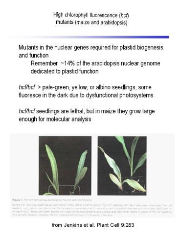
High chlorophyll fluorescence (hcf) mutants
(maize and arabidopsis)
Mutants in the nuclear genes required for plastid
biogenesis and function Remember 14 of the
arabidopsis nuclear genome dedicated to plastid
function hcf/hcf pale-green, yellow, or
albino seedlings some fluoresce in the dark due
to dysfunctional photosystems hcf/hcf seedlings
are lethal, but in maize they grow large enough
for molecular analysis
from Jenkins et al. Plant Cell 9283
2
Plant organelle RNA processing
Polycistronic transcripts undergo extensive,
complex processing prior to translation e.g.
psbB operon in maize, encoding subunits of two
different plastid protein complexes psbB / psbH
/ petB / petD The nuclear mutation crp1 disrupts
processing of the polycistronic message, PETA,
PETB and PETD protein accumulation Models Failur
e to accumulate monocistronic petD transcripts
results in failure to translate petD because the
petD initiation codon is buried in secondary
structure in the dicistronic petB / petD
transcript failure to translate PETD may
destabilize PETB and result in failure to
translate PETA CRP1 is bi-functional and directs
translation of PETA and PETD, independent of its
role in processing the petB-petD intercistronic
spacer
3
Pentatricopeptide repeat (PPR) proteins Lurin et
al. Plant Cell 162089
One of the largest multigene families in plants
(441 members in arabidopsis vs 7 in
humans) Primarily plastid or mitochondrial
targeted Implicated in post-transcriptional RNA
metabolism through single gene/mutant analysis
e.g. crp1 locus in maize necessary for plastid
petB / petD RNA processing e. g.
restorer-of-fertility loci for CMS in petunia,
radish and rice all influence processing or
stability of mitochondrial CMS gene transcripts
and encode PPR proteins Why so many? (? Guides
for RNA editing) How do they function? (?RNA
binding adaptors that recruit enzymatic protein
complexes to act on RNA in a site-specific
manner)
4
Pentatricopeptide repeat (PPR) proteins Lurin et
al. Plant Cell 162089
Figure 3. Motif Structure of Arabidopsis PPR
Proteins (35 amino acid repeats). Typical
structures of proteins from each of the principal
subfamilies and subgroups are shown. The
structures are purely indicative, and the number
and even order of repeats can vary in individual
proteins. The number of proteins falling into
each subgroup is shown.
5
Pentatricopeptide repeat (PPR) proteins Lurin et
al. Plant Cell 162089
We assume that the putative superhelix formed by
tandemly repeated PPR motifs forms a
sequence-specific RNA binding surface either
alone (A) or in the presence of an additional
factor (B). The resulting protein-RNA complex
recruits one or more other transfactors to a
specific site on the RNA target (in this case an
endonuclease). We assume that in most cases the
catalytic site is in the partner protein for the
DYW class of PPR proteins, it may lie in the
C-terminal domain itself.
6
RNA editing genetic analysis defines a
trans-acting factor
Figure 1 Characterization of crr4 mutants. a,
Schematic model of NDH function. The NDH complex
functions in electron transport from the stromal
reducing pool, NADPH and reduced ferredoxin (Fd),
to the plastoquinone pool (PQ). PQ reduction in
the dark depends on NDH activity and is detected
in the transient rise of chlorophyll fluorescence
after illumination with actinic light (AL)6. PSI,
photosystem I PSII, photosystem II. b, Analysis
of the transient increase in chlorophyll
fluorescence after turning off AL. The bottom
curve indicates a typical trace of chlorophyll
fluorescence in the wild type (WT). Leaves were
exposed to AL (50 µmol photons m-2 s-1) for
5 min. AL was turned off and the subsequent
transient rise in fluorescence ascribed to NDH
activity was monitored by chlorophyll
fluorimetry. Insets are magnified traces from the
boxed area. crr4-X CRR4, crr4 alleles
transformed by the wild-type genomic CRR4
sequence. ML, measuring light SF, saturating
flash. c, Immunoblot analysis of thylakoid
proteins. Immunodetection of an NDH subunit
(NdhH) and photosystem II (PsbO). The lanes were
loaded with proteins corresponding to 0.5 µg of
chlorophyll for PsbO and tenfold the proteins for
NdhH (100) and a series of dilutions as
indicated.
from Kotera et al. Nature 433326
7
RNA editing genetic analysis identifies a
trans-acting factor
Figure 2 Structure of CRR4. a, Schematic
alignment of CRR4 and CRR2. The relationship
between PPR motifs (boxed) and PCMP motifs (bars
AH) is shown. PCMP motifs are labeled on the
basis of the original assignment10 except for the
E motif12. The sites of mutation in four crr4
alleles are indicated. White boxes are the
putative plastid targeting signals. The
C-terminal 15-amino-acid motif is indicated by a
bar labelled with an asterisk. b, Alignment of
eleven PPR motifs present in CRR4. Amino acids
conserved more than 60 are boxed in black.
Conserved similar amino acids are shaded. The
points of amino acid alteration in three alleles
are highlighted by red letters. A pair of
antiparallel -helixes, predicted from the
similarity with TPR motif11, is shown by
underlines.
from Kotera et al. Nature 433326
8
RNA editing genetic analysis identifies a
trans-acting factor
Figure 3 Analysis of RNA editing in the ndhD
initiation codon. a, Direct sequencing of RTPCR
products containing the ndhD initiation codon.
The psaC and ndhD region is shown schematically.
RNA editing sites are indicated. The restriction
enzyme NlaIII cleaves cDNA derived from edited
molecules. The editing site is so distal in the
transcripts that cDNA was sequenced on the
complementary strand. b, Semi-quantitative
analysis of the extent of RNA editing. RTPCR
products were digested with NlaIII. Fragments
originating from edited and unedited RNA
molecules are indicated. WT, wild type.
from Kotera et al. Nature 433326


