Definitions - PowerPoint PPT Presentation
1 / 97
Title: Definitions
1
Definitions
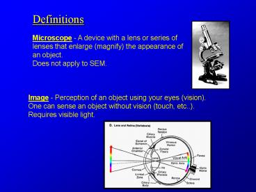
Microscope - A device with a lens or series of
lenses that enlarge (magnify) the appearance of
an object. Does not apply to SEM.
Image - Perception of an object using your eyes
(vision). One can sense an object without vision
(touch, etc..). Requires visible light.
2
Lens - A lens is an optical component which is
used to focus beams of radiation. Lenses for
light are usually made of a transparent material,
whereas non-uniform electromagnetic fields are
used as lens for electrons.
Curved glass or mirror for Visible light
convex
concave
Concave surface of metal (e.g. satellite
dish) Radio waves
3
Concave mirror or Fresnel lens Heat
Solenoid (electromagnetic fields that can be
varied) Subatomic particles (electrons, protons,
positrons)
4
Magnification - The ratio between image size to
the object size. Can be varied by changing the
distance between the object and the final lens
(of the eye) or by inserting a second lens
between the two.
Resolution - The point at which two or more
objects can be distinguished as separate
individual objects.
5
History First record of using glass lens for
magnification was by Al Hazen, an Muslim
scientist from what is now known as Iran, in the
10 and 11th century. He performed the bulk of
his studies and work in Spain. His treatise on
the colors of the sunset were translated to
Latin. He contradicted Ptolemy's and Euclid's
theory of vision that objects are seen by rays of
light emanating from the eyes. According to Al
Hazen, the rays originate from the object of
vision and not in the eye. Because of his
extensive research on vision, he has been
considered by many as the father of modern
optics.
http//home.att.net/mleary/alhazen.htm
6
Antonie van Leeuwenhoek
15th century on - Studies done with glass
magnifiers to study objects in detail mostly as a
curiosity by non-scientists - Antonie van
Leeuwenhoek (dry goods merchant) described three
shapes of bacterial cells using his simple,
single lens microscope (glass bead in metal
holder). Was probably influenced by Robert Hooke.
7
Instructions on making A Leeuwenhoek microscope
http//www.mindspring.com/7Ealshinn/Leeuwenhoekpl
ans.html
8
Robert Hooke In 1665, Hooke described cork and
other microorganisms in a drop of water. First
to produce a book on microscopical observations.
Made several modifications creating a compound
microscope. Few improvements were made to the
light microscope until the 19th century.
9
By mid-19th century, became evident that
theoretical resolution limits of light were
reached. Above a magnification of 1,500
resolution lost. The image can be magnified,
but blurred (empty magnification).
10
Wavelength - the distance between peaks of the
waveform
In 1870, Ernst Abbe derived mathematical
expression for resolution of microscope Resoluti
on is limited to approx. 1/2 the wavelength of
illuminating source
11
Resolution ? ½ ?
12
Shortest visible wavelength - Blue light has a
wavelength of 0.47 um. Resolution max 0.2 um
(200 nm) Cannot go beyond this even with better
optics. Solution? Use illumination of shorter
wavelength
Antone de Broglie (1924) Theory of wave nature
of electrons Hermann Busch (1924) axial magnetic
fields refract electrons Electron optics
13
- 1935 - Max Knoll demonstrates the theory of the
scanning electron microscope
1938 - First scanning electron microscope
produced by von Ardenne
Knoll and Ruska 1986 Nobel Prize winners
1939 - Ruska and von Borries, working for Siemens
produce the first commercially available EM
14
- 1939 - First EM built in North America by James
Hillier and Albert Prebus at the University of
Toronto
Dr. Ladd
Dr. Prebus
15
(No Transcript)
16
Light vs Electron Microscope
17
Transmission Electron Microscopy
l ? ( 150 / V )1/2 Angstroms
Substituting 200 eV for V gives l a of 0.87
Angstroms
For a beam of 100 KeV we get a wavelength of
0.0389 and a theoretical resolution of 0.0195
Angstroms! But in actuality most TEMs will only
have an actual resolution 2.4 Angstroms at 100KeV
18
Electron Sources
Similar in design to a tungsten filament
19
Electron Sources
Filament Current (Heating Current) Current
running through the emitter Beam
Current Current generated by the emitter
20
Transmission Electron Microscope Optical
instrument in that it uses a lens to form an
image Scanning Electron Microscope Not an
optical instrument (no image forming lens) but
uses electron optics. Probe forming-Signal
detecting device.
21
Electron Optics
Electrostatic lens
Must have very clean and high vacuum environment
to avoid arcing across plates
22
Electromagnetic Lens
23
The strength of the magnetic field is determined
by the number of wraps of the wire and the amount
of current passing through the wire. A value of
zero current (weak lens) would have an infinitely
long focal length while a large amount of current
(strong lens) would have a short focal length.
24
A TEM image is made up of nonscattered electrons
(which strike the screen) and scattered electrons
which do not and therefore appear as a dark area
on the screen
25
Some of the scattered
electrons will only be partially scattered and
thus will reach the screen in an inappropriate
position giving a false signal and thus
contributing to a degradation of the image.
These forward scattered electrons can be
eliminated by placing an aperture beneath the
specimen.
26
The design of an electromagnetic lens results in
a very strong lens with a very short focal length
thus requiring that the specimen lie within the
lens itself along with an aperture to stop the
highly scattered electrons
27
The simplest way to correct for chromatic
aberration is to use illumination of a single
wavelength! This is accomplished in an EM by
having a very stable acceleration voltage. If the
e velocity is stable the illumination source is
monochromatic
28
Goals of Specimen Preparation Observe specimen
near natural state as possible. Preservation
of as many features as possible. Avoid artifacts
(changes, loss or additional information) Render
specimen stable for examination in environment of
TEM.
29
Problems
TEM not widely used by biologists until 1950s
Considerations for TEM- High vacuum Support of
sample Intense heat from beam Depth of electron
penetration
Considerations for SEM- High vacuum Size of
specimen Localized elevated temperatures Capable
of emitting signal Conductive
30
Specimen preparation
Stabilization - Fixation and dehydration. Embeddin
g in resin for TEM Surface Preparation -
cleaning and/or exposure of new surface for SEM.
Cutting specimen to ultra-thin sections for
TEM Mounting - specimen on stub (SEM) or grid
(TEM) Staining with heavy metals for image
contrast (TEM)
31
Basic factors affecting chemical fixation
pH (Isoelectric point) Total ionic strength of
reagents Osmolarity Temperature Length of
fixation Method of application of fixative
32
Common Buffers used in Fixation
Sodium Cacodylate Effective range is 6.4 - 7.4.
Lacks phosphates that could interfere with
cytochemical studies. Incompatible with uranyl
salts and should be rinsed out thoroughly if
planning to do "en-bloc" staining with UA. Can
add Calcium and Magnesium without precipitation.
Used extensively with animal tissues. Contains
arsenic, which is toxic. Avoid contact with acids
to avoid production of arsenic gas.
33
Fixation
A process which is used to preserve (fix) the
structure of freshly killed material in a state
that most closely resembles the structure and/or
composition of the original living state.
Chemical crosslinking - coagulative/noncoagulative
- Coagulative original killing agents
(alcohols, Farmers, FAA, Bouins) Low
pH Unbuffered Coagulates cellular components -
like frying an egg. - Non Coagulative
Formaldehyde, Glutaraldehyde, Osmium Tetroxide
34
Glutaraldehyde
- Glutaric acid dialdehyde, a 5 Carbon dialdehyde,
is the most widely applied fixative in both
scanning and transmission electron microscopy. - Most highly cross-linking of all the aldehydes.
GTA fixation is irreversible. - In TEM, buffered GTA has the reputation of
providing the best ultrastructural preservation
in the widest variety of tissue types of any
known chemical fixative.
35
Osmium Tetroxide (OsO4)
- A non-polar tetrahedral molecule with a molecular
weight of 254 and solubility water and a variety
of organic compounds. - Its principle utility is its ability to stabilize
and stain lipids- preferentially unsaturated
fatty acids - Although it is widely used in preparative schemes
for SEM, this must be due at least in part to
arbitrary whole-cloth adoption of TEM fixation
schemes for SEM. Except for cases where lipid
retention is essential, - the aforementioned qualities of this compound
have much less to offer the area of SEM. - Commercially available as a coarse yellow
crystalline material packaged in glass ampoules
sealed under inert gas. Similarly packaged
aqueous solutions are also available.
36
Tissue
Standard Preparation
TEM
SEM
Chem. Fixation
Cryo Fixation
Chem. Fixation
Cryo Fixation
Rinse/store
Substitution
Rinse/store
En bloc staining
Cryo- sectioning
Dehydration
Dehydration
Dehydration
Drying
Resin infiltration
Mounting
Sectioning
Coating
Post staining
37
Embedding and Sectioning
- Requirements for cutting any material into thin
slices - Support - biologicals tend to be soft. Inducing
hardness in them gives them the mechanical
support needed for sectioning. - Accomplished by lowering temperature (freezing)
or infiltration with some material that can be
hardened. - Plasticity - resiliency as opposed to
brittleness.
38
Embedding and Sectioning
- Cryosectioning
- Commonly done for light microscopy.
- ie hospital operating room biopsies.
- Rapid.
- Preservation is usually sufficient for a rapid
diagnosis. - Overall resolution is low.
- Ultrathin cryosectioning
- Technically demanding
- Requires expensive specialized equipment
- Ultrastructural preservation often poor due to
freezing artifact. - Usually done only when tissue cannot be exposed
to chemical fixatives...as in some
immunolabeling, analytical work.
39
Embedding and Sectioning
- TEM Embedment
- Tissue infiltrated with a resin which is
polymerized by heat, chemicals, or U.V. - Provides support to section infiltrated tissue to
about 40 nm minimum. - Infiltration is limited...specimens can be no
more than a few mm thick. - The required thinness of the sample and the
friction during cutting limits the section size
to about 1 mm2 maximum.
40
Embedding and Sectioning
- Infiltration
- In resin/solvent mixture in increasing
concentration - Ethanol/resin or acetone resin often used
- Propylene oxide/resin is most effective
- When 100 resin is reached, it should be changed
twice to insure that all solvent is removed - Polymerization
- Thermal - 50-70 C, depending on resin mix
- U.V. - usually done to avoid heat
- of polymerization. Often done at low temp.
41
Embedding and Sectioning
- Ultramicrotomy
- Mechanical Advance
- Thermal Advance
- Ultramicrotome Knives
- Diamond - 1.5 - 6mm cutting edge
- Latta-Hartmann (glass) - 6mm cutting edge (1mm
useable) - Both use water to support and lubricate the
section as it is cut (decreases friction)
42
Embedding and Sectioning
- Making a glass knife
- Use of a glass knifemaker to score a 1" glass
square
43
Embedding and Sectioning
A scored 1" glass square (top) and the resultant
glass knife
Making the water trough Tape or plastic
a) Cutting edge b) Knife angle (45o) c) Corner d)
Shelf
44
Setting up the Microtome
Block face
Sample Block
Knife edge
Glass Knife
45
Embedding and Sectioning
- Section Thickness
- Ideally, sections should be in the 55 - 60 nm
range. - This allows for enough stain uptake for contrast,
and maximum resolution (limited in the TEM by
specimen-induced chromatic aberration). - Determined by interference colors.
- Maximum thickness should not exceed 85 - 90 nm
(light gold). - Thickness can sometimes be reduced by one color
range by flattening sections - smooths out
compression to a limited extent. Toluene,
xylene, chloroform, heat.
46
Section Mounting
- A 200m grid has 60 open area a 400m grid only
40 - Thin-bar grids...more fragile, more expensive.
- Ultrathin sections can be supported on a bare
grid of no greater than 200m.
- Commonly used TEM grid types
47
Picking up sections
Mesh grids
Eyelash tool
Slot grids
48
Section Mounting
An ultrathin section on a 50m support filmed grid
at 200X mag.
49
(No Transcript)
50
(No Transcript)
51
Mitochondria
52
(No Transcript)
53
Golgi (Dictyosome)
54
Post-Staining
- Normally done, even if en bloc staining (ie
uranyl acetate) has been done. - Uranyl acetate - 0.5 - 2 aqueous or saturated
ethanolic or methanolic - Lead citrate - several formulations (Venable and
Coggeshell Reynolds) mostly using lead nitrate
chelated with sodium citrate. - Adequate rinsing between and after staining is
essential to prevent post-stain contamination. - Particular care must be used to exclude CO2 to
inhibit lead carbonate formation - black
cannonballs.
55
Contrast
- Transmission Electron Microscopy
- Heavy metals commonly used for contrasting in
TEM uranium, lead, osmium, ruthenium,
molybdenum, gold, silver. - It is the differential adsorption of various
heavy metals to tissue components that produces
the electron image of biological thin-sectioned
materials. - The image may be composed of areas ranging from
completely black to completely white with all
ranges of grey in between. - Images with mostly pure blacks and whites are
"contrasty" images, while those containing mainly
greys are "flat images.
56
One Stage and Two Stage Replicas
57
(No Transcript)
58
Cryo-Preservation
Why?
- Structures are ephemeral or events are rapid.
- Structures are fixative sensitive.
- Removal of water changes topography/morphology
Rapid arrest of cellular components Avoidance of
artifacts from chemical fixation Preserves sample
in hydrated state Maintains structural and
cellular integrity Cellular domains are
maintained (e.g. IMPs) Ice crystal formation can
be avoided Sublimation (etching) used to remove
excess water
59
Slam Freeze Gentleman Jim
60
Endoplasmic reticulum
Fungus hyphal tip
61
Freeze-fracture
Sample is rapidly frozen, fractured and a
replica is made. Etching (sublimation) of the
sample may be used to expose features.
62
Fracture surface with frozen blade
63
Fracture Plane Views
Fracture
Cut
64
(No Transcript)
65
TEM of Golgi sections and freeze fracture
66
Freeze-fractured replica of the alga Dunaniella
67
Immunolocalization for EM
Using immunoglobulin molecules as tags for select
proteins and carbohydrates. Visualized by using
colloidal gold or enzyme reactions
Leishmania megasome labeled with 10nm gold
68
Generic TEM Immunolabeling Protocol
Fixation Glutaraldehyde only Dehydration Embe
dding in methacrylate resin (e.g. LR White,
Lowicryl, or Quetol) Section Immunolabel Option
al - Post-stain
69
Immunolabeling Sections
- Float grids on blocking solution - Incubate in
primary antibody - Wash thoroughly with
buffer to remove unbound antibody
70
Incubate with secondary - different size gold
Note steric hinderence at asterisk
71
10 nm and 5 nm gold
72
Ferritin enhanced labeling
Silver enhanced
73
Freeze fracture immunolabeled
Negative stain Immunolabeled
74
Conventional SEM
Specimen at high vacuum requires sample
fixation and dehydration or freezing. Charging
is minimized by coating sample with metal or
carbon or lowering the operating kV.
75
Variable Pressure Scanning Electron Microscope
- - Vacuum in the sample chamber can range from
high vacuum (lt 10-6 Pascals) up to 3,000 Pa. - - Gas in the sample chamber allows uncoated and
unfixed samples to be imaged. - Detectors used at higher pressures are
backscatter or special secondary detectors. - - Moisture on the sample can be controlled by
cooling/heating stage and water injection system.
76
Applications
Live centipede
Bacteria on rock
77
Fresh moss with liquid water
78
Skyscan 1072 Micro-CT X-Ray Tomography Scanner
79
Sasov and van Dyck, 1998, J. Microsc.
Object is rotated 180 degrees. Images captured
at one degree increments. Reconstructions done
on aligned images to create volume data.
80
Oak Ridge Natl Lab
81
(No Transcript)
82
(No Transcript)
83
(No Transcript)
84
(No Transcript)
85
Credit where credit is due The University of
Georgia EM course (www.uga.edu/caur./teaching.htm)
Dr. Mark Farmer (markfarmer_at_cb.uga.edu) Dr.
John Shields (jshields_at_cb.uga.edu) And then
there is me. Paul Linser (pjl_at_whitney.ufl.edu)
86
Semi-intact mosquito larva with intact digestive,
nervous, and tracheal systems
- Boudko, Moroz, Linser, Trimarchi, Smith, and
Harvey 2001, J. E. B. 204, 691-9
87
Gross Anatomy
88
Integument and associated tissues
dorsal
ventral
Heart
Anopheles gambiae
Fat body
Ventral nerve cord
89
Tracheal Trunk and HeartDorsal view
Tracheal Trunk
Heart
Pericardial cells
90
Ventral Nerve Cord
5HT
FMRFamide-like
91
Gastric Caeca
Cardia
tracheae
AMG
92
Muscle, nerve, trachea and gut epithelium
93
Muscle
Tracheae
Muscle
94
Immunolabeling by antibody to V-ATPase
- Zhuang et al., J. Exp. Biol. 202, 2449-2460
95
V-ATPase Portasomes
96
Anterior midgut basal membrane is decorated by
portasomes
- Zhuang et al., J. Exp.
- Biol. 202, 2449-2460
97
AMG to PMG Transition
Clark et al.,Tissue Cell 37457-68, 2005


