
Retina Examination PowerPoint PPT Presentations
All Time
Recommended

Eye is made up of Iris, Pupil, Cornea and Retina. The retina is an extremely thin tissue that lines the inside of the back of the eye. It is the light-sensitive portion of the eye. Light from the objects we are looking at, enters the eye. Cornea and the eye lens focus the light image onto the retina. Human eye works like a camera, light striking the retina causes a complex biochemical change within certain layers of the retina and this, in turn, stimulates an electrical response within other layers of the retina.
| PowerPoint PPT presentation | free to download
Fundoscopic Examination Window to the blood vessels Normal Ocular Fundus Fundoscopic Examination Hypertensive retinopathy Diabetic retinopathy Bacterial endocarditis ...
| PowerPoint PPT presentation | free to download
Eyes Inspection Visual Acuity Visual Fields Pupillary Response Fundoscopic Exam Eye Examination Inspection Eyes Visual Acuity Eyes Extraocular Movements Convergence ...
| PowerPoint PPT presentation | free to download

Collie eye anomaly. Syndrome in the. Collie. Shetland Sheepdog. Border Collie. Australian Shepherd. Three types of defects. Chorioretinal dysplasia 'Go normals' ...
| PowerPoint PPT presentation | free to view

Week 3 Visual Pathway and Visual field defects a= bjerrum area B=papillomacular bundle causing central and ceccocentral scotomas C=nasal bundle horazontal raphe wedge ...
| PowerPoint PPT presentation | free to download
High Success Retina Surgery. What Retina Surgeons Do and When to See One. Dry Macular Degeneration: What It Is and What You Should Know.
| PowerPoint PPT presentation | free to download

Title: Nursing Board Review Author: morpheus Last modified by: Myk Created Date: 10/19/2004 2:47:18 AM Document presentation format: On-screen Show
| PowerPoint PPT presentation | free to download

Retinal Vein Occlusions Morphology CRVO BRVO Hemispheric VO Hemicentral VO Papillophlebitis Macular BRVO CENTRAL RETINAL VEIN OCCLUSION The actual mechanisms ...
| PowerPoint PPT presentation | free to download

The intravenous fluorescein angiogram pattern of an ischemic central retinal ... with the extent of capillary nonperfusion on the fluorescein angiogram. ...
| PowerPoint PPT presentation | free to view

EXAMINATION OF THE CRANIAL NERVES OLFACTORY NERVE (I) Test with alcowipes, coffee etc. Unilateral anosmia may be significant Bilateral anosmia: commonest cause viral ...
| PowerPoint PPT presentation | free to view

1. Eye Health – Caring for your Retina. 2. Diabetes and the prevention of Retinal Problems. 3. Retina Problems can be Associated with Age. 4. Latest Advances in Retina Treatments for Vision Loss.
| PowerPoint PPT presentation | free to download
1. Take care of your eyes and they will last forever 2. When our general health affects our eyes 3. Ophthalmology Examinations are Important
| PowerPoint PPT presentation | free to download

Retina is a layer of tissue at the back of the eye, which helps to see images focused on it by the cornea and lens. Retinal Detachment is an eye disorder, wherein the retina gets separated from the underlying layer of blood vessels, which supplies oxygen and other nutrients to it.
| PowerPoint PPT presentation | free to download

The global retinal imaging devices market is growing at a CAGR of 5.93% and is expected to reach $9322.89 million, during the forecast period of 2023-2032.
| PowerPoint PPT presentation | free to download

7/29/09. Slit Lamp Examination - IC. 1. INSTRUCTION COURSE ON SLIT LAMP EXAMINATION ... Prerequisites Preferably a dark room and slit lamp biomicroscope ...
| PowerPoint PPT presentation | free to view

DACH (dachshund) Has coiled-coil domain, probably dimerises; shown to interact with eya. ... DACH: dachshund = mouse/human Dach1, Dach2. A conserved team at ...
| PowerPoint PPT presentation | free to view
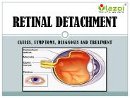
Retina is a layer of tissue at the back of the eye, which helps to see images focused on it by the cornea and lens. Retinal Detachment is an eye disorder, wherein the retina gets separated from the underlying layer of blood vessels, which supplies oxygen and other nutrients to it. When the retina gets detached, the supply of oxygen and nutrients are stopped. If the condition is left untreated, it may even lead to a complete vision loss and blindness. To know more visit here: www.lazoi.com
| PowerPoint PPT presentation | free to download

HRT (Heildelberg Retina Thomograph) is widely used for imaging and following ... Optimizers: controlled random search (CRS), Powell. ...
| PowerPoint PPT presentation | free to download
While speaking in particular to the ophthalmologists, they are very much comfortable in using the software as there results in a diagrammatic representation of retina and even goni. All the arithmetic calculations are looked over by the system. Checkout Our Ophthal Software Presentation and If You have any queries then let us know at http://triocorporation.in
| PowerPoint PPT presentation | free to download

... lies in front of the pigment epithelium that lines the back of ... cones, each with a distinct pigment that is most sensitive to ... type of rod pigment ...
| PowerPoint PPT presentation | free to view

The retina is a very complex part of the eye, having a very complicated functioning. Thus, even the tiniest of change seen in the eye must be reported to the best retina specialist in Mumbai at the earliest!
| PowerPoint PPT presentation | free to download

You need to get in touch with the best retina specialist in Mumbai if you are facing any kinds of eye problems to detect if there is a problem in any part of the retina, so as to treat it at the earliest.
| PowerPoint PPT presentation | free to download

Dr. Ron P Gallemore, MD, California, CA, Los Angeles, Ophthalmology , 321 North Larchmont Boulevard 1020, (323) 464-9393. Get a FREE Background Report on Dr. R
| PowerPoint PPT presentation | free to download

Retina specialist in Mumbai say that it is one of the more common misdiagnosis as one has to be aware of the minute details. They say that CRVO tends to develop disc edemas and twisted blood veins in the eye, which don’t occur in diabetic retinopathy. Hence, both these conditions require a unique treatment approach.
| PowerPoint PPT presentation | free to download

Retinal detachment is a severe condition that can result in complete vision loss, which is why if any symptoms are experienced, must be immediately reported to the best retina specialist in Mumbai.
| PowerPoint PPT presentation | free to download

required vision examinations and vision screenings for children: current research william t. reynolds, o.d. richmond, kentucky joel n. zaba, m.a., o.d.
| PowerPoint PPT presentation | free to view

Retinal detachment is a severe condition that can result in complete vision loss, which is why if any symptoms are experienced, must be immediately reported to the best retina specialist in Mumbai.
| PowerPoint PPT presentation | free to download

Retinal detachment is a severe condition that can result in complete vision loss, which is why if any symptoms are experienced, must be immediately reported to the best retina specialist in Mumbai.
| PowerPoint PPT presentation | free to download

While the best retina specialist in Mumbai will always strive to identify the precise condition of every patient’s eye, there are instances when one condition may be misinterpreted for another due to minute differences.
| PowerPoint PPT presentation | free to download

Office(JEC 7010): 518-276-8067, Lab(JEC 6308): 518-276-8207, Fax: 518-276-8715 Email: roysam@ecse.rpi.edu, Web: http://www.ecse.rpi.edu/~roysam Output Size: 1200 x 820
| PowerPoint PPT presentation | free to download

Dr. Delia Cabrera dcabrera2@med.miami.edu. Value Added to CenSSIS. Optomap ... Special thanks to Dr. Delia Cabrera, from Bascom Palmer Eye Institute at the ...
| PowerPoint PPT presentation | free to view

Uniformed Services University of the Health Science, Bethesda, MD ... Dilated fundus examination: Healthy Retina. No signs of any intraocular lens displacement ...
| PowerPoint PPT presentation | free to view
Direct Ophthalmoscopy By Thomas Anders Brevik What is it used for? Examine the retina and its structures Also known as funduscopy (examination of the fundus) Turning ...
| PowerPoint PPT presentation | free to download

Jan Jyoti Super Speciality Eye Hospital has established itself as the Best Eye Hospital in Jabalpur, offering comprehensive and advanced eye care services. With a commitment to excellence, the hospital provides state-of-the-art treatments and personalized care, ensuring optimal outcomes for patients. Advanced Eye Care Services At Jan Jyoti, patients receive the Best Eye Care in Jabalpur, encompassing a wide range of services: Retina Care: Expert Retina specialists near me offer precise diagnosis and treatment for retinal disorders, utilizing cutting-edge technology.
| PowerPoint PPT presentation | free to download

PEDIATRIC OPHTHALMOLOGY UNITS/ RETINA UNITS/ GLAUCOMA UNITS IN EYE DEPARTMENTS ... Strengthening of Pediatric Ophthalmology Units. Retina Units. Glaucoma Units. ...
| PowerPoint PPT presentation | free to view

Board Review Ophthalmology By Stacey Singer-Leshinsky R-PAC Vision Image focused by cornea and lens onto retina Light absorbed by photoreceptors in retina (rods and ...
| PowerPoint PPT presentation | free to download

Human eye has various important parts like Cornea, Pupil, Iris, Lens and Retina. The macula is located in the center of the retina, the light-sensitive tissue at the back of the eye. The retina instantly converts light, or an image, into electrical impulses.
| PowerPoint PPT presentation | free to download

* * * * * * * * * * * * * * * * * * * * * * * * * * * * * * * * Physiology of Photoreceptors Vertebrate ... Receptive Fields in the Retina Two ...
| PowerPoint PPT presentation | free to download

Fundus appearance. Prognosis / visual outcome. Treatment. CRAO ... Fundus ... Havreb et al. Fundus changes in central retinal artery occlusion. Retina. ...
| PowerPoint PPT presentation | free to view

The case history will determine if examination of the eyes is ... Arcus Senilis. Eye. Examination. Inspection: Iris & Pupil. Meiosis. Mydriasis. Anisocoria ...
| PowerPoint PPT presentation | free to view
Mental status Kamal Shemisa PGY3 Internal Medicine UHCMC Objectives Definitions Neurological Examination Clinical Presentation Diagnostic Evaluation Encephalopathy vs ...
| PowerPoint PPT presentation | free to download

Sharp Sight is the best eye hospital in East Delhi. You can visit Sharp Sight any day for your eye examination.
| PowerPoint PPT presentation | free to download

Sharp Sight is the best eye hospital in East Delhi. You can visit Sharp Sight any day for your eye examination.
| PowerPoint PPT presentation | free to download

Sharp Sight is the best eye hospital in East Delhi. You can visit Sharp Sight any day for your eye examination.
| PowerPoint PPT presentation | free to download

Astigmatism is a condition that occurs when light that enters the eye is not focused evenly onto the retina.
| PowerPoint PPT presentation | free to download

DEFINITION The clinical electro-oculogram is an electrophysiological test of function of the outer retina and retinal pigment epithelium in which the change in the ...
| PowerPoint PPT presentation | free to download

Maintaining good eye health is crucial for overall well-being. Regular eye checkups at Jan Jyoti Super Speciality Eye Hospital can help detect early signs of eye conditions, ensure timely treatment, and preserve your vision for years to come. Whether you have perfect eyesight or use corrective lenses, periodic eye examinations are essential for everyone.
| PowerPoint PPT presentation | free to download

Universal scholar with a sense for the practical. Ophthalmoscope for examining the retina ... Call for Applications. Helmholtz - Fellowship Programs ... Ul. ...
| PowerPoint PPT presentation | free to download

-Prevent Blindness America Diabetic Eye Disease Diabetic Retinopathy Damage to blood ... Smoking Pregnancy Take Home Annual Dilated Eye Examination ...
| PowerPoint PPT presentation | free to view

Ears and Eyes By Donald Hudson, D.O., FACEP/ACOEP Ears External Examination (Auricle and Mastoid) Size Shape Symmetry Color Position Tenderness Odor Ears Otoscope ...
| PowerPoint PPT presentation | free to view

The conjunctiva is clear and colorless except when blood vessels are dilated. ... A normal fluoresein angiogram. Detached Retina ...
| PowerPoint PPT presentation | free to view

Are you aware of diabetic retinopathy? If not, you're in the right place to learn how it can be a serious sight-threatening complication associated with diabetes. This eye disease, caused by high blood sugar levels, damages the back of the eye (retina). The main issue with this condition is that it often shows no warning signs until it has already progressed. Therefore, it's crucial to seek timely treatment before the condition worsens. Through this presentation, we aim to spread awareness, as we believe that awareness can make a significant difference.
| PowerPoint PPT presentation | free to download

direct inspection of the fundus. Examination. Media Opacities. Corneal edema: ... Normal pupillary reactions and fundi. Chronic Visual Loss. Chapter 3. Introduction: ...
| PowerPoint PPT presentation | free to view

Explore how high blood pressure affects eye health and causes risks like damage to the optic nerve and retina. Learn the signs that necessitate the requirement of an eye exam and treatment from the best eye doctors Indio at Acuity Optical.
| PowerPoint PPT presentation | free to download

in premature babies peripheral retina is not vascularised at birth ... Toxocariasis (nematode infection) from exposure to puppies ...
| PowerPoint PPT presentation | free to view

During the first two decades of disease, nearly all patients with type 1 ... Puberty. Pregnancy. Lack of appropriate ophthalmic examination ...
| PowerPoint PPT presentation | free to view