Micrograph PowerPoint PPT Presentations
All Time
Recommended
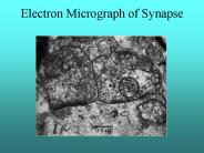
Electron Micrograph of Synapse Brainstem Limbic System Cerebral Cortex Lobes of the Brain Thalamus The LGN LGN to V1 Electron Micrograph of Synapse Brainstem Limbic ...
| PowerPoint PPT presentation | free to download
... used to thoroughly numb the area. We don't do surgery unless you are numb! ... Once numb, the soft part of the cancer is scraped away. Mohs Micrographic Surgery ...
| PowerPoint PPT presentation | free to view
Wet Mount PT: 2010A Micrographs 1-9 Evaluate images 1 - 9 separately as if each is a representative field from an individual patient (some s include both low and ...
| PowerPoint PPT presentation | free to download
Wet Mount PT: 2011A Micrographs 1-9 Evaluate images 1 - 9 separately as if each is a representative field (low and high power) from an individual patient
| PowerPoint PPT presentation | free to download
The simplest micrograph uses a 250mL conical flask, upside down, and resting over the eyepiece. A piece of frosted (translucent) plastic or tracing paper covers the "bottom" of the flask and is held in place by a rubber band. This forms a viewing screen. The other type requires the construction of a light weight wooden box mounted on legs. A hole is cut in the bottom of the box so that the box will fit over the eye piece of the microscope. A hole is cut in the top of the box and a piece of frosted glass is used to cover the hole. This becomes the viewing screen.
| PowerPoint PPT presentation | free to view
MOHS MICROGRAPHIC SURGERY (MMS) Hayleigh Gordon Histology BMS 1 What is MOH s ? Mohs surgery or MMS is a specialised technique enabling the surgeon to remove a skin ...
| PowerPoint PPT presentation | free to view
2003 EIPBN MicroGraph Contest. Magnification (3'x4' image): Instrument (Make and Model): Submitted by: Affiliation: Micrograph Title: Description: ...
| PowerPoint PPT presentation | free to view
micro & nano - graph Contest. Magnification: 33X. Submitted by: Richard Stallcup ... Electrochemical etching of a tungsten wire produces an extremely fine probe and ...
| PowerPoint PPT presentation | free to view
MOHS MICROGRAPHIC SURGERY: A HISTORICAL PERSPECTIVE Patricia Ting, BSc, Anatoli Freiman, MD Division of Dermatology, McGill University Health Centre, Montreal, Canada
| PowerPoint PPT presentation | free to download
Neurons interpret electrochemical signals from multiple ... 'Canny' Edge Detection Algorithm Outline: Remove white noise. Apply an edge-detecting operator. ...
| PowerPoint PPT presentation | free to view
Low-power scanning electron micrograph of osteoporotic bone. architecture in the 3rd lumbar vertebra of a 71 yr old woman ... marrow and other cells removed to ...
| PowerPoint PPT presentation | free to download
The X-Ray structure of horse heart cytochrome. myoglobin = single subunit ... Figure 10-15 The heme group and its environment in the unliganded a chain of human Hb. ...
| PowerPoint PPT presentation | free to view
Figure 9-36 Molecular formula for iron-protoporphyrin IX (heme). Page 313 ... Figure 10-15 The heme group and its environment in the unliganded a chain of human Hb. ...
| PowerPoint PPT presentation | free to view
Figure 22-37 Electron microscopy based image of E. coli F1F0 ATPase. Page 828 ... unit at low ATP concentration as observed by fluorescence microscopy. Page 833 ...
| PowerPoint PPT presentation | free to view
????????????? ???????????????? ?????????? ????-??????????: ... ???????????????? ?????? ?????????: ???????? ??????- ???????? ????????. ????? ... HREM micrograph of ...
| PowerPoint PPT presentation | free to view
a Light micrograph (phase-contrast process) b Light micrograph (Normarski process) c Transmission electron micrograph, thin section. d Scanning electron micrograph ...
| PowerPoint PPT presentation | free to view
View electron micrograph of bacteria. ... [light micrograph, gram stain] ... This electron micrograph shows the 9 2 pattern of microtubules in a single ...
| PowerPoint PPT presentation | free to view
Any Question? SEM micrograph of nanocomposite of Carbon nano ...
| PowerPoint PPT presentation | free to view
Nucleus Micrograph. Nuclear Pores ... Endoplasmic Reticulum Micrograph. 47. Ribosomes. Small structures on which proteins are made. ... Golgi Complex Micrograph ...
| PowerPoint PPT presentation | free to view
Micrograph Data Set: using electron micrographs for students to ... Micrographs are measured at team benches ... Micrograph data set. Student Errors Found ...
| PowerPoint PPT presentation | free to view
Micrograph courtesy of PNNL. 8/11/09. 28. 8/11/09. 29. NiAl Conversion. 15 minutes. 3 hours ... Micrograph courtesy of PNNL. Micrograph courtesy of PNNL ...
| PowerPoint PPT presentation | free to view
FST 305 GENERAL MICROBIOLOGY By Prof. Olusola Oyewole And Dr. Olusegun Obadina Viral Structure Drawing Electron Micrograph Drawing Electron Micrograph Helical ...
| PowerPoint PPT presentation | free to view
??? ????: ?????? ??? ????????? ??????? ???????? ?????? . ????, ??'? ???? ?????? ??????. ... TEM micrograph. ZnO 1.5% in LDPE. Effect of ZnO loading on AF ...
| PowerPoint PPT presentation | free to view
The major intracellular compartments of an animal cell ... INTRACELLULAR COMPARTMENT. An electron micrograph. Development of plastids ...
| PowerPoint PPT presentation | free to download
LM micrographs of striated muscle. Low power EM micrograph. High power EM micrograph ... Arrangement of the myosin molecules within the filament (250-350 ...
| PowerPoint PPT presentation | free to view
Microscopy and Microanalysis for phase transformation studies, ... FIM micrograph showing , ', and ... Other FIM micrographs indicated fine distribution of and ...
| PowerPoint PPT presentation | free to view
... are visible in electron micrographs. Adapted from Fig. 4.6, ... Micrograph of. brass (a Cu-Zn alloy) 0.75mm. Optical Microscopy. crystallographic planes ...
| PowerPoint PPT presentation | free to view
Testicular Histology. Scanning Electron Micrograph. Light Micrograph. Seminiferous Tubules ... What is the function of the spermatogonia? Clonal expansion ...
| PowerPoint PPT presentation | free to view
Thin sensitive/readout layer for compact calorimeter design ... CERN GDD Group electron-micrograph of GEM foil. CERN GDD Group electron-micrograph ...
| PowerPoint PPT presentation | free to download
FST 305 GENERAL MICROBIOLOGY By Prof. Olusola Oyewole And Dr. Olusegun Obadina Viral Structure Drawing Electron Micrograph Drawing Electron Micrograph Helical ...
| PowerPoint PPT presentation | free to view
( D) Electron micrograph of a microtubule assembled from purified tubulin and ... Micrograph showing Kranz anatomy in maize, a C4 plant. ...
| PowerPoint PPT presentation | free to view
( B) Fluorescence micrograph of pollen tubes growing through a pistil. The scanning electron micrograph at left shows the surface of a stigma. ...
| PowerPoint PPT presentation | free to view
Using a microscope, Robert Hooke ... Scanning electron micrograph of cilia. Transmission electron microscope (TEM) Transmission electron micrograph of cilia ...
| PowerPoint PPT presentation | free to view
Electron Micrograph of. RER and Mitochondria. Mitochondria. The site of Cellular Respiration ... Electron Micrograph. Chloroplast. Found only in plant cells ...
| PowerPoint PPT presentation | free to view
Refers to the appearance of this organelle in electron micrographs ... Electron micrograph of sections: Flagellum. Basal body. Basal body ...
| PowerPoint PPT presentation | free to view
Preparations for prototype construction. Test GEM 'chamber' designed at UTA ... CERN GDD Group electron-micrograph of GEM foil. CERN GDD Group electron-micrograph ...
| PowerPoint PPT presentation | free to download
Optical micrograph ... Optical micrograph. Av=61.8dB, fc=493.9KHz, PM=52.14 for fu=539MHz ... Optical micrograph. Simulated: td=1.6ns, Max Op Freq=500MHz ...
| PowerPoint PPT presentation | free to view
Dr. Dev Shah is qualified dermatolist in the North West London. He is the Skin Surgeon specialising in Mohs Micrographic Surgery.
| PowerPoint PPT presentation | free to download
... AISI 304 stainless steel plate (as welded) and micrograph of the root region ... Higher magnification micrographs showing absence of cracks in weld centreline ...
| PowerPoint PPT presentation | free to view
Rough Endoplasmic Reticulum Fluorescent Micrographs of ER TEMs of RER F37 Free And Membrane- Bound R-somes The signal hypothesis: ...
| PowerPoint PPT presentation | free to download
a measure of how greatly a substance slows the velocity of light ... Transmission Electron Micrograph. Scanning Electron Micrograph. Newer Techniques in Microscopy ...
| PowerPoint PPT presentation | free to view
Fig. 1. Grain structure micrographs of the AZ31 alloy after ... and (b) ECAP at 200oC (TEM micrograph). Fig. 2. Grain size versus annealing temperature ...
| PowerPoint PPT presentation | free to download
SEM micrograph of 2 m thick film grown onto -Al2O3 substrate and annealed in dry ... SEM micrograph of the 1 m thick films grown by the sequential-deposition method ...
| PowerPoint PPT presentation | free to view
This is an SEM micrograph of an extracellular matrix forming around a cocci type ... This SEM micrograph shows an Extracellular Polysaccharide Substance (EPS) ...
| PowerPoint PPT presentation | free to view
Micrograph of a Utah Electrode Array (UEA) with 100 equal length probes. ... Optical micrographs of initial Au coil samples on polyimide substrates with a ...
| PowerPoint PPT presentation | free to view
NANOFIBERS FOR APPLICATIONS IN MEMS, NANOTECHNOLOGY, BIOTECHNOLOGY AND MEDICINE Electrospinning of nanofibers Electron micrograph of 300 nm polybenzimidazole ...
| PowerPoint PPT presentation | free to view
Hopeless situation? A VLSI MOS transistor An analog chip layout: ... Electron Micrograph of a Real Neuron Mahowald & Mead s ... single-chip solution includes ...
| PowerPoint PPT presentation | free to download
EM radiation is produced by accelerating charges. example ... Electron micrograph. light micrograph. X-rays. f ~ 1016-1020 Hz ~ 10-8 -10-12 M. Atomic dimensions ...
| PowerPoint PPT presentation | free to view
... patterns and TEM micrograph which characterize the nanocrystalline ... The synthesis produces also electroactive carbon nanoflasks, as seen in the micrograph. ...
| PowerPoint PPT presentation | free to view
Nagy7, A. Kereszturi7 and H. Hargitai7 ... 3Geological Survey of Canada, ... CL micrograph and spectrum of apatite. Reference material. based on Goetze, 1999 ...
| PowerPoint PPT presentation | free to download
... of lambda and T4 tail tubes Micrographs of T4 tail tubes containing 5 key proteins Gp29 fiber is visible on contracted tails and whole phage T4 ...
| PowerPoint PPT presentation | free to view
Tools of the Laboratory: The Methods for Studying Microorganisms * Insert figure 3.25 Scanning micrographs Wet mounts and hanging drop mounts allow examination of ...
| PowerPoint PPT presentation | free to download
vertical-cavity surface-emitting laser (semiconductor laser device, diode laser) ... Etched Mesa VCSEL(electron micrograph) Schematic Etched Mesa VCSEL. ...
| PowerPoint PPT presentation | free to view
A SEM micrograph of a stoma of a pea plant: ... A TEM micrograph of a chloroplast: From: http://www.botany.uwc.ac.za/ecotree/leaves.htm#top ...
| PowerPoint PPT presentation | free to view
THE EVOLUTION OF MICROSCOPES Early Electron Microscope Modern Electron Microscope Scanning Electron Microscope SEM Micrograph Transmission Electron Microscope TEM ...
| PowerPoint PPT presentation | free to view
Supplemental figure Figure S1. Morphology of starch granules of endosperm from WT and RSR1-overexpressing lines. Scanning electron micrographs of endosperm cross ...
| PowerPoint PPT presentation | free to download


