INVESTIGATION OF FIBROBLAST BIOMECHANICS BY MICROPIPETTE ASPIRATION TECHNIQUE - PowerPoint PPT Presentation
1 / 12
Title:
INVESTIGATION OF FIBROBLAST BIOMECHANICS BY MICROPIPETTE ASPIRATION TECHNIQUE
Description:
INVESTIGATION OF FIBROBLAST BIOMECHANICS BY MICROPIPETTE ASPIRATION TECHNIQUE S.P. Krechetov 1, V.V. Smol aninov 2 Mechanical Engineering Research Institute, – PowerPoint PPT presentation
Number of Views:233
Avg rating:3.0/5.0
Title: INVESTIGATION OF FIBROBLAST BIOMECHANICS BY MICROPIPETTE ASPIRATION TECHNIQUE
1
INVESTIGATION OF FIBROBLAST BIOMECHANICS BY
MICROPIPETTE ASPIRATION TECHNIQUE
- S.P. Krechetov 1, V.V. Smol?aninov 2
- Mechanical Engineering Research Institute,
Russian Academy of Sciences, - Moscow, 117334, Russia
- E-mail 1- ksp__at_mail.ru, 2- smolian_at_mail.ru
2
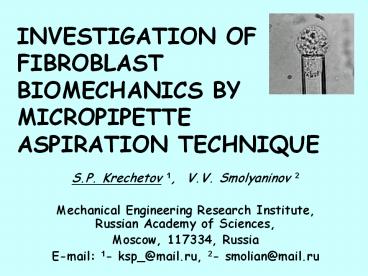
Typical view of fibroblast before and after
aspiration in micropipette
2
10 mm
10 mm
before aspiration
after aspiration
3
3
Experimental assembly for cells aspiration
- 1 - syringe, 2 leveling vessel, 3
underpressure vessel, 4 - three-way cock, 5 -
water manometer, 6 micropipette holding head,
7 micropipette, 8 measurements cell, 9 -
fibroblast, 10 microscope, 11 light source.
4
Measurements cameras
4
A thin camera for adhesive cells. B thick
camera for non adhesive cells. 1 cover glass, 2
stainless still strip, 3 - object-plate, 4
filling port, 5 paraffin fixation.
5
5
Fibroblast geometry before and after aspiration
6
6
Fibroblast membrane isotropic deformation model
? ? 0k?(S/S0-1)
? ? 0
before aspiration
after aspiration
7
Functions describe fibroblast deformation geometry
7
MATLAB 6.5
8
Membrane tension as function of cell geometry
8
9
NMF and L-cells membrane parameters in step
aspiration experiments
9
Pout-Pin, d, l?y, D0?X0
? ? 0k?(S/S0-1)
d 11 mm, D0 19 mm ? X0 1.7
10
NMF and L-cells membrane parameters in similar
aspiration conditions experiments
10
Pout-Pin, d, l?y, D0?X0
? ? 0k?(S/S0-1)
3
2
1
1 - Pout-Pin 2 cm H2O, d 9 mm, D0 14.3
mm
2 - Pout-Pin 10 cm H2O, d 5mm, D0 14.8 mm
3 - Pout-Pin 14 cm H2O, d 5 mm, D0
14.7 mm
11
Fibroblast elasticity changes under influence of
different factors
11
Factor Testing cells and factor levels Elasticity changes description Elasticity changes description Elasticity changes description
Factor Testing cells and factor levels Elasticity changes parameter calculation Elasticity changes parameter value Elasticity changes direction
Cell transformation ??? ? L-cells lL-cells/lNMF 0.57 ? 0.08 (4) Increase
Temperature increasing L-cells, 25?C ? 37?C l37?/l25? 1.38 ? 0.43 (8) Decrease
Increasing of macroergic phosphates L-cells, ATP 10 mM lATP/lN 0.82 ? 0.06 (4) Increase
Microfilaments destruction L-cells, cytochalasin 10 mg/ml lcytochalasin /lN 1.49 ? 0.16 (8) Decrease
Microtubules destruction L-cells, colcemid 0.1 mg/ml lcolcemid/lN 1.01 ? 0.14 (6) Non significant
Microfilaments and microtubules destruction L-cells, cytochalasin 10 mg/ml and colcemid 0.1 mg/ml lcytochalasincolcemide/lN 1.73 ? 0.24 (7) Decrease
Fetal calf serum (FCS) NMF Eagles medium ? 10 FCS lwithout FCS./lN 1.04 ? 0.01 (4) Non significant
Serum albumin (HSA) L-cells Eagles medium ? 1 HSA lwithout HAS./lHSA. 1.20 ? 0.17 (8) Non significant
Glutaric aldehyde (GA) treatment L-cells, 0.001 GA lGA/lN 0.92 ? 0.11 (8) Increase
Glutaric aldehyde (GA) treatment L-cells, 0.005 GA lGA/lN 0.53 ? 0.13 (7) Increase
Glutaric aldehyde (GA) treatment L-cells, 0.01 GA lGA/lN 0.28 ? 0.09 (6) Increase
12
Conclusions
12
- Isotropic cortical biomechanical fibroblast model
agree with aspiration technique experimental
data. - Transformed fibroblasts are more rigid in
relation to normal fibroblasts. Elastisity
coefficient of cortical structures (membrane) is
10-25?10-3 N/m for normal fibroblasts and
40-100?10-3 N/m for transformed. Membrane tension
of strainless fibroblast is below 1?10-3 N/m for
normal fibroblasts and near 1?10-3 N/m for
transformed. - Fibroblast rigidity increase after glutaric
aldehyde treatment and adding ATP in culture,
decrease after cytochalasin treatment and at
temperature rise. No significant changes in cell
tonus observed after colcemide in culture medium,
removal from it fetal calf serum or replace fetal
calf serum by human serum albumin.


