
Cell Division - PowerPoint PPT Presentation
1 / 14
Title:
Cell Division
Description:
Cell Division Background Info What, specifically does DNA really code for? Codes for Amino Acids that assemble to make a protein What is the normal ... – PowerPoint PPT presentation
Number of Views:84
Avg rating:3.0/5.0
Title: Cell Division
1
Cell Division
2
Background Info
- What, specifically does DNA really code for?
- Codes for Amino Acids that assemble to make a
protein - What is the normal appearance of DNA under a
microscope? - CHROMATIN (not chromosomes) LOOKS LIKE
SPAGHETTI, OR A HAIRBALL - What is the point of DNA tightening together into
chromosomes before cell division? - For organization before Cell Division occurs
3
- How many chromosomes are in a human cell?
- 46 chromosomes 23 from mommee, 23 from daddio
- There are only 23 types of chromosomes
- Mitosis making identical copies of a nucleus
- Q What part of cell division does the definition
of mitosis leave out? - Among other things (1) the doubling of
organelles, and (2) the cell actually splitting
4
Mitosis creates two genetically identical
nuclei (2.5.5)(Mitosis is only part of the
cells life cycle)
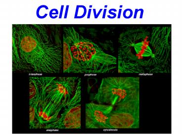
- Cell Cycle (life of a cell) consists of
(2.5.1) - Interphase
- Mitosis
- Prophase
- Metaphase
- Anaphase
- Telophase
- Cytokinesis division of the cytoplasm
5
- B. Interphase the longest phase an active
growth period when many biochemical reactions are
occurring (2.5.3) - 3 stages of interphase
- G1 formation of new organelles (ribosomes,
mitochondria, etc) transcription and translation
of proteins - DNA transcription making mRNA from DNA
- Translation making proteins from mRNA
- S DNA Replication
- G2 spindle fibers form chromosomes condense
6
Interphase
NOTE diagrams are misleading because the DNA is
NOT really coiled up yet into chromosomes (which
is why transcription and DNA synthesis can occur)
A a C C
G1
B b d
D
MOM
DAD
Homologous pair of chromosomes
At this point, is the cell haploid or
diploid? Its diploid- there is a copy of each
chromosome from both parents.
7
- Haploid or diploid?
- Still diploid- now with sister chromatids.
- Heterozygous or homozygous?
- Heterozygous for genes A, B, D
- Homozygous (dominant) for gene C.
S
A A a a C C
C C
B B b b d d
D D
G2 microtubules join to form the
spindle
CENTROMERE (the region joining 2 sister
chromatids)
2 chromatids (still one chromosome)
8
C. Mitosis (2.5.4, 2.5.5)
- Prophase - Supercoiling of chromosomes
C C d d
a a b b
A A
C C D D
B B
Centriole (made of microtubules)
- Nuclear membrane breaks down
- Centrioles move to the poles
- (No centrioles in most plant cells animals
only!) - Spindle microtubules are growing
- DNA begins to coil into chromosomes
9
Metaphase spindle microtubules attach to
centromeres at kinetochores chromosomes line
up at the equator on separate spindle
microtubules nuclear membrane has broken down
kinetochore
A A
B B
a a b b
(pole)
C C D D
(pole)
centriole
C C d d
equator
10
Anaphase centromeres split and chromosomes move
toward the poles, pulled by spindle microtubules
sister chromosomes move to opposite poles
A B a b
C D C d
A B a b C D
C d
11
ANAPHASE!!!!!
Spindle Fibers
Cytoplasm
A B A B
D d
A B A B
D d
Centrioles
Centrioles
12
Telophase chromosomes reach the poles spindle
microtubules break down cytokinesis begins
during late telophase
PLANT ANIMAL
-forms a cell plate -forms a cleavage
furrow
-Nuclear envelope reforms
A a
C C
D B b
d
a C A
C b D B
d
13
- D. Cytokinesis division of the cytoplasm to
form two (identical) cells - II. Differences in mitosis between plant and
animal cells - -plant cells do not have centrioles in animal
cells they are found at each pole during mitosis - -plant cells cannot form cleavage furrows and so
they form cell plates
14
- III. Reasons for mitosis (2.5.6)
- Growth
- Embryonic development
- Repairing damaged tissue
- Asexual reproduction
- IV. Tumor formation (2.5.2)
- Repeated, uncontrolled cell division
- Can happen in any organ, and can spread to other
parts of the body (metastasis) - When it causes disease it is considered a cancer
Recommended
CrystalGraphics Presentations


