FINGERTIP INJURIES - PowerPoint PPT Presentation
1 / 24
Title:
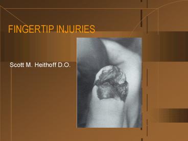
FINGERTIP INJURIES
Description:
FINGERTIP INJURIES Scott M. Heithoff D.O. INTRODUCTION Fingertip injuries are defined as those distal to the insertion of the flexor and extensor tendons Primary goal ... – PowerPoint PPT presentation
Number of Views:1924
Avg rating:5.0/5.0
Title: FINGERTIP INJURIES
1
FINGERTIP INJURIES
- Scott M. Heithoff D.O.
2
INTRODUCTION
- Fingertip injuries are defined as those distal to
the insertion of the flexor and extensor tendons - Primary goal of treatment is a painless fingertip
with durable and sensate skin - Methods of treatment include healing by secondary
intention, skin grafting, shortening of the bone
and primary closure, and coverage with local or
regional flaps
3
ANATOMY OF THE FINGERTIP
- The skin covering the pulp of the finger is very
durable and has a thick epidermis with deep
papillary ridges - The thick skin beneath the distal free edge of
the nail plate is called the hyponychium - The pulp consists of fibrofatty tissue that is
stabilized from the dermis to the periosteum of
the distal phalanx
4
ANATOMY OF THE FINGERTIP
- The nail complex, or perionychium, includes the
nail plate, the nail bed, and the surrounding
skin on the dorsum of the fingertip (paronychium) - The dorsal skin over the nail fold is called the
nail wall - The distal margin of the nail wall, which adheres
to the nail plate, is called the eponychium
5
ANATOMY OF THE FINGERTIP
- The nail bed is adherent to the very thin
periosteum over the distal two thirds of the
distal phalanx and consists of the sterile and
germinal matrices - The germinal matrix is located proximally and
forms the ventral floor of the nail fold. - The lunula is the distal margin of the germinal
matrix - The sterile matrix is the portion of the nail bed
distal to the lunula and is adherent to the nail
plate
6
EVALUATION
- History and mechanism of the injury
- Patient factors age, gender, handedness,
occupation, and history of previous hand injuries - Function of flexor and extensor tendons
- Radiographs
- IV antibiotics and tetanus prophylaxis
7
EVALUATION
- Anesthesia is best obtained via a digital nerve
block - A bloodless field is essential, and this can be
facilitated with the use of a penrose drain - It is important to know whether there is loss of
skin pulp tissue and the amount of loss, is there
bone exposed, and is there an injury to the nail
bed - Treatment is dependant on the above information
8
EVALUATION
- It is also important to determine the angle of
amputation - A,B Volar oblique
- C Transverse
- D Dorsal oblique
9
SOFT-TISSUE LOSS WITHOUT EXPOSED BONE
- There are two treatment options for this injury
- Skin graft
- Healing by secondary intention
- Most agree that for smaller wounds (lt1 cm2)
should be treated nonoperatively by the open
method
10
SOFT-TISSUE LOSS WITHOUT EXPOSED BONE
- Treatment via open technique
- Complete healing takes 3 - 5 weeks and occurs by
wound contraction and epithelialization - 7 - 10 days after the injury, the patient is
instructed to begin soaking the finger in a warm
water-peroxide solution once a day and to apply a
light bandage and fingertip protector
11
SOFT-TISSUE LOSS WITHOUT EXPOSED BONE
- Skin grafting should be considered for larger
wounds (gt1 cm2) - Skin grafts applied to the palmer surface of the
fingertip should be full thickness because they
contract less, are more durable and less tender,
and achieve better sensibility than split grafts - Grafts should be taken from a hairless area, such
as the hip or volar surface of the wrist
12
SOFT-TISSUE LOSS WITH EXPOSED BONE
- When bone is exposed, satisfactory soft-tissue
coverage must be obtained. - Treatment by the open method after the bone has
been shortened below the level of the skin may
result in a good outcome, by is associated with
an unacceptable incidence of nail-plate
deformities - Treatments include revision amputation, local
flaps, or regional flaps
13
SOFT-TISSUE LOSS WITHOUT EXPOSED BONE - REVISION
AMPUTATION
- Shortening and primary closure of fingertip
amputations is indicated in adults of any age
when not enough sterile matrix (lt5mm) remains to
produce an adherent, stable nail - The remaining nail matrix must be ablated, and
this can be accessed by reflecting the nail wall
proximally - If the flexor and extensor tendons cannot be
preserved, the DIP should be disarticulated,
traction applied to the tendons, then transected
14
SOFT-TISSUE LOSS WITHOUT EXPOSED BONE - LOCAL
FLAPS
- Defined as a flap in which the transferred tissue
is confined to the injured digit, with at least
one side of the flap adjacent to the defect - Advantages can be used in patients of any age,
they preserve length, the donor defect does not
require a skin graft, and the transposed tissue
is similar in quality, texture, and color to that
of the recipient site - Types V-Y flap (Kleinert) and Kutler flap
15
SOFT-TISSUE LOSS WITHOUT EXPOSED BONE - LOCAL
FLAPS V-Y FLAP
- This flap is ideal for transverse or dorsal
oblique amputations - The critical value is whether enough palmer
tissue is available for distal advancement - Patients usually have near normal sensation and
good restoration of contour and padding
16
SOFT-TISSUE LOSS WITHOUT EXPOSED BONE - LOCAL
FLAPS KUTLER
- This flap is most appropriate for distal
transverse amputations - The disadvantage of this technique is that the
flaps are small and may be difficult to advance
17
SOFT-TISSUE LOSS WITHOUT EXPOSED BONE - REGIONAL
FLAPS
- The two most commonly used regional flaps are
cross-finger flap and the thenar flap - Used for amputations that are volar oblique or
too proximal to allow a local flap - The main disadvantage is it involves a two stage
procedure - Contraindicated in patients with osteophytes or
arthritis of the involved digits and in patients
with systemic conditions such as RA, diabetes,
and vasospastic disorders
18
SOFT-TISSUE LOSS WITHOUT EXPOSED BONE - REGIONAL
FLAPS CROSS-FINGER FLAP
- The standard cross-finger flap is a rectangle
over the middle phalanx of the donor digit, with
the hinge side adjacent to the injured finger - A full-thickness skin graft from the groin or
elsewhere is applied to the donor defect - Flap division is performed 12-14 days after the
initial procedure
19
NAIL-BED INJURIES
- Spectrum of injuries
- Subungual hematomas
- Simple and complex lacerations
- Avulsions of matrix tissue
- It is important that the nail bed be repaired
with great attention to detail in order to
restore function and prevent annoying and
unsightly deformities
20
NAIL-BED INJURIES - SUBUNGUAL HEMATOMAS
- Decompression of a subungual hematoma should be
performed to relieve pain if it involves lt 50 of
the area of the nail - This can be done with a heated paper clip or 18
gauge needle - For larger subungual hematomas, the nail plate
should be removed to repair the nail bed
21
NAIL-BED INJURIES - LACERATIONS
- Lacerations are repaired after the nail plate has
been removed - The nail plate is carefully separated from the
nail matrix with a Freer elevator - The wound is then irrigated and debrided, taking
care that all matrix tissue is retained - Fractures of the distal phalanx can usually be
stabilized by suturing the skin of the lateral
nail folds and the nail bed
22
NAIL-BED INJURIES - LACERATIONS
- Lacerations of the skin and lateral nail folds
should be repaired with 5-0 nylon suture - The nail bed is meticulously approximated with
absorbable 6-0 chromic or plain gut suture - If the laceration extends into the germinal
matrix, the nail wall should be reflected
proximally by making an incision on each side of
it, extending from the eponychium - After repair, the nail plate should be placed
back into the nail fold to prevent scar formation
23
SUMMARY
- For the treatment of fingertip injuries, the
decision making process should proceed from the
simpler techniques to the more complicated - When no bone is exposed, the open method is ideal
for small or moderate sized wounds, and skin
grafting should be considered for larger wounds - Distal transverse and dorsal oblique amputations
with bone exposure can be treated with local
advancement flaps
24
SUMMARY
- More proximal and volar oblique amputations can
be managed with a regional flap to preserve
length if enough sterile matrix remains for a
stable nail - Shortening and primary closure can be used for
amputations not amendable to other methods of
treatment


