The Plague - PowerPoint PPT Presentation
Title:
The Plague
Description:
Over a course of time, Yersinia Pestis aquired additional plasmids and pathogenicity islands that broaden its range of hosts and change its lifestyle entirely. – PowerPoint PPT presentation
Number of Views:499
Avg rating:3.0/5.0
Title: The Plague
1
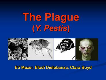
The Plague (Y. Pestis)
Eti Mezei, Elodi Dielubanza, Clara Boyd
2
Overview - 3 Types of Plague
Bubonic Pneumonic Septicemic
- Most common form - Responsible for most plague epidemics - Generally secondary complication of bubonic plague though can be a primary infection - Diagnosis is more difficult - Easily communicable - Generally secondary complication of bubonic plague though can be a primary infection - Poses the greatest burden to the human system of the 3 types
All types of plague are caused by the
gram-negative bacteria Yersinia Pestis
3
Epidemiology Historical Modern Aspects
Reservoirs, Vectors Transmission World
History Discoveries Modern Plague Trends in the
USA
4
Reservoirs Vectors
Reservoirs Vectors Incidental Hosts
Urban and domestic rats Ground squirrels Rock squirrels Prairie dogs Deer mice Field mice Gerbils Voles Chipmunks Marmots Guinea pigs Kangaroo Rats Xenopsylla cheopis (the oriental rat flea nearly worldwide in moderate climates) Oropsylla montanus (United States) Nosopsyllus fasciatus (nearly worldwide in temperate climates) Xenopsylla brasiliensis (Africa, India, South America) Xenopsylla astia (Indonesia and Southeast Asia) Xenopsylla vexabilis (Pacific Islands) Humans Domestic and feral cats Dogs Lagomorphs (rabbits and hares) Coyotes Camels Goats Deer Antelope
5
Modes of Transmission
- Fleabite
- Inhalation of salivary droplets of infected
humans or cats - Cats scratch
- Ingestion of the bacillus
- Contact with infected body fluids
6
Transmission Type of Plague
Bubonic Septicemic Pneumonic
1? Y. Pestis enters the lymphatic system through the skin Bite from infected flea Direct inoculation of Y. Pestis into bloodstream Bite from infected flea Scratch from infected cat Contact with Infected Fluids Direct inhalation or ingestion of Y.Pestis Inhaling droplets from cough of infected human/cat Eating infected animal Inhaling Y. Pestis in the lab
2? ----------------- As a complication of bubonic or 1? pneumonic plague Via hematogenous spread, as a complication of bubonic or septicemic plague
7
Transmission Cycles
8
Plague and World History
- 1320 B.C. -- 1st mention of plague is in the
Bible Philistines stole the ark of the covenant
from the Israelites and plague ensued - the Lords hand was heavy upon the people
of Ashdod and its vicinity. He brought
devastation upon them and afflicted them with
tumors. And rats appeared in their land, and
death and destruction were throughout the
cityThe Lords hand was upon that city,
throwing it into great panic. He afflicted both
young and old with and out break of tumors in the
groin.
9
Plague and World History
- 541-700 A.D. 1st Pandemic, Justinian Plague
- Began in Pelusium Egypt and
- spread to the middle East
- Europe with estimated population
- losses of 50-60 in N. Africa,
- Europe S. Asia.
- A.D. 558-654 The 2nd through 11th epidemics
occurred - in 8-12 year cycles.
10
Plague and World History
- 1346-1666 2nd Pandemic, Black Death
- Plague believed to have entered Europe along
trade - routes from central Asia by fleas in
bundles of imported fur. - 1347-1351 1st 5-year epidemic killed an
estimated 17-28 million people, 30-40 of
Europes population at the time. - Epidemics continued throughout the period in 2-5
year cycles until 1480 and then with less
frequency until late 17th century. - The plague stimulated significant
- advance in medical practice including
- the beginnings of clinical research,
- reorganization of hospitals.
A Plague Doctor
NOTE other epidemics existing at the time of the
1st and 2nd Plague pandemics may have contributed
to depopulation figures
11
Plague and World History
- 1855 3rd Pandemic, China
- Began in the Yünnan province and troops from
the war in that area helped to spread plague down
the southern coast to Hong Kong and Canton by
1894 and Bombay by1898. - By 1900 steamships had helped to spread the
disease to Africa, Austrailia, Europe Hawaii,
India, Japan, the Middle East, the Phillipines,
the U.S, and S. America. By 1903, 1 million
people/year were dying of plague in India.
Mukden, China, 1910-11 Workers in a plague
hospital hose off an autopsy table with
carbolic spray
12
Plague and World History
- 1855 3rd Pandemic, China (contd)
- Stable enzootic foci were established on every
inhabited continent except Austrailia and
outbreaks continue until today though greatly
reduced because of advances in public health
practices and drugs. - Significantly, it is during the 3rd Pandemic that
the bacterium responsible for plague as well as
its vectors and reservoirs were identified.
13
History of Yersina Pestis
- First discovered in 1894 during the Hong Kong
Plague by two independent investigators
(Alexandre Yersin and Shibasaburo Kitasato)
within days of each other. - Kitasato was initially credited for the discovery
but Yersins bacillus proved to be the infecting
bacillus. - Yersin drained buboes of deceased plague victims
and identified the bacillus with microscopy. - Confirmed the involvement of the bacillus in
infection by injecting healthy rodents with
infected lymph isolates. Healthy animals
developed plague symptoms and dissection showed
blood and organs to be filled with the isolated
bacillus.
14
Y
- Nomenclature the plague bacillus has had four
name changes. - Classifications
- Bacterium Pestis until 1900
- Bacillus Pestis until 1923
- Pasturella Pestis (after Yersins mentor)
- Yersinia Pestis in 1970 until present day
15
Identification of Vectors and Reservoirs
Yersin also confirmed the Black rat (Rattus
rattus) as the reservoir after local
suspicion implicated its involvement in the
transmission of the plague. In 1897 during
the Indian outbreak, Paul- Louis Simond and
Masanori Ogata identified the oriental rat flea
(Xenopsylla Cheopis) as the transmission
vector by exposing healthy rodents to fleas
collected from the corpses of rats which had
died recently from plague.
16
Early Prevention and Treatment Methods
- Yersin isolated serum from horses immunized with
the bacillus for treatment of human plague in
1896. - W.M Haffkine created and used an effective
preventative vaccine for the Plague containing
killed bacteria during the Manchurian outbreak
(1910). - L.T Wu characterized pneumonic plague during the
1910 Manchurian outbreak and helped to institute
protective measures against aerosol spread of the
disease.
17
Modern Plague
- For most of the 20th century the occurrence of
plague has sharply decreased though not
disappeared. Plague remains an enzootic infection
of rats, ground squirrels and other rodents in
every inhabited continent except Austrailia. - Why has Plague decreased so sharply?
- The WHO credits public health prevention
protocols and the development of effective
antibiotics.
18
Modern Plague
- WHO reports 1000-3000 cases of plague worldwide
annually - (avg.1700cases/year for the last 50 years)
(conservative estimate) - Plague is largely underreported in countries with
limited surveillance and laboratory capabilities. - Modern plague is primarily occurrent in rural
areas with poor sanitation and large rodent
populations. - Urban cases are increasingly rare. Los
Angeles1924 was the last U.S urban outbreak.
19
Modern Plague - Distribution
largest enzootic foci are in the Southwestern
Pacific U.S and the former USSR.
20
Modern Plague Cases Reported to WHO 1954-1997
21
Modern Plague - Timeline
- 1900 - Plague officially arrives in the U.S.
Infected corpse of Chinese Laborer found in San
Francisco hotel basement. - 1924 - Los Angeles. 33 cases, 31 fatal. 1st case,
Mexican American male, misdiagnosed with STD,
family and neighbors contract plague and die with
in two weeks. Many die before public health
measures were taken. - 1967-72 - Vietnam. Defoliation, disturbance of
the ecosystem and economic losses during the war
contributed to outbreak. Singular contributor to
the 1967 jump in worldwide plague vases. - 1992 - Arizona. 1 confirmed case, 1 fatality.
Misdiagnosis of pneumonia. Post-mortem lab tests
confirmed Y. Pestis. Family cat identified as
source.
22
Timeline Contd
1994 India. Bubonic plague begins when
rats arrive because of stockpiles of relief
grain. Pneumonic form shortly follows. 5150
suspected pneumonic and bubonic cases from 26
states. 163 confirmed with serology, 53 confirmed
deaths, 300 suspected. Mass panic 600,00 flee
Surat including 110 plague cases.
23
Timeline Contd
- 1997
- Zambia- Jan. Namwala region 90 cases of bubonic
plague, 22 fatal. - Heavy rains drove rats into inhabited regions.
- Mozambique - Aug. Tete province. 115 reported
cases of bubonic. - No reported deaths.
- Malawi - Oct. Southern region, 43 reported
cases, 17 of which were seropostive. gt60 cases
were children. No reported deaths. - Mozambique - Nov. August outbreak continues and
extends to 225 cases. - No reported deaths.
- 1999
- Namibia - May Northwest region, 39 reported
cases, 8 deaths. - Malawi - July Reported cases in 22 villages, 74
suspected cases total. - No reported deaths.
24
Timeline Contd
- 2001
- Zambia - Mar. Nyanje and Petauke regions 436
suspected cases, 11 deaths. Y. Pestis postively
identified. - 2002
- India - Feb. Hat Koti village. 16 cases of
pneumonic plague including 4 deaths. Cases linked
to one villager. - Malawi- June. Nsanje region and 26 nearby
villages. 71 reported cases of bubonic plague. - 2003
- New York - Nov. 2 cases. Couple contracts plague
in Santa Fe, NM and travels to New York. Neither
case fatal but man required double foot
amputation.
25
Plague Patient Returns HomeNEW YORK, Feb. 10,
2003
A New Mexico man who was hospitalized in New York
City for more than three months with bubonic
plague left the hospital to fly home on Monday, a
spokesman said. John Tull left Beth Israel
Medical Center at about 7 a.m., hospital
spokesman Mike Quane said. Tull was admitted to
Beth Israel on Nov. 5. Tull, whose feet were
amputated due to extensive tissue damage, will
begin physical therapy in Albuquerque, N.M.,
Quane said. Disease investigators believe Tull
and his wife, Lucinda Marker, contracted plague
from infected fleas on their Santa Fe, N.M.,
ranch. They became ill after arriving in New York
on Nov. 1 for vacation.
John Tull at the Beth Israel Hospital in New York
City with his wife Lucinda Marker.
CBSNEWS.com
26
Trends in Human Plague in the U.S.
- From 1899-1926 Plague in the U.S. was an urban
epidemic involving domesticated rats with cases
most prevalent in California and Hawaii. - Late 1940s saw a jump in infection in the
Southwestern states which remain hotspots for
infection. - Static annual infection levels from 1925-1964
(avg.lt2 cases/year). 1965 saw increases that
would carry into the 80s.
27
Trends in Human Plague in the U.S
- Seasonal distribution 1926-1979 81.5 of plague
cases occurred in May-September. Prior to 1926
most were Sept.-Oct. - 1970-1980
- 53 cases ?
- 59 were lt 20 years old.
- Similar age distribution from 1926-1969
- Racial dist. 1970-79 35 of cases were in
Indians (1.4 Indians per 100,00 compared to
0.1non-Indians).
28
Simplified Pathogenesis (Focus on Bubonic Plague)
29
Pathogenesis
Flea feeds on Y. Pestis-infected blood Y.
Pestis enters fleas midgut multiplies
logarithmically Clump of Y.Pestis fibronous
material forms in the midgut, blocking fleas
proventriculus During next meal, blood cannot
enter the midgut flea gets very hungry Flea
bites vigorously regurgitates the contents of
its midgut into the next wound
Blocked Flea
30
Pathogenesis
ENTRY Flea bite -- regurgitation of
blood containing Y. Pestis ( 25,000 -100,000
organisms) into interstitial space of
subcutaneous tissue
DISSEMINATION Superficial lymph vessels drain
skin subcutaneous tissue --Y. Pestis enters
the lymphatic system
31
Pathogenesis
DISSEMINATION Y. Pestis migrates through
lymph vessels to regional lymph node
Massive inflammatory response obliterates
underlying lymph node architecture -- leads to
painful swellings (Buboes)
32
Pathogenesis
HEMATOGENOUS DISSEMINATION Y. Pestis may enter
the blood stream at the lymph node, and can then
travel to other organs (liver, spleen). ( 2?
Septicemic Plague)
HEMATOGENOUS DISSEMINATION Once in the
bloodstream, Y. Pestis also has the
opportunity to enter the lungs. (2? Pneumonic
Plague)
33
Molecular Biology of Yersinia Pestis
Bacteriological Characteristics Evolution Biovars
Genome Molecular Pathogenesis
Proteins/Virulence Factors/ Immunology
34
Bacteriological Characteristics
- Gram negative
- Non-motile
- Enterobacteriacae
- Non-spore forming coccobacillus
- Facultative anaerobe
- Obligate parasite
35
Bacteriological Characteristics
- .5-.8 uM in diameter
- 1-3 uM long
- Grows optimally at 28 C and a pH of 7.2-7.6
- Bacterial cell wall
- Protein Envelope (F1)
36
Evolution of Y. Pestis
- There are 11 species of Yersinia
- 3 pathogenic species of Yersinia
- Yersinia Pseudotuberculosis - enteropathogen
- Yersinia Enterocolitis - enteropathogen
- Yersinia Pestis - systemic pathogen
- Y. Pestis evolved from Y.pseudotuberculosis
1500-4000 years ago - 90 chromosomal DNA relatedness
- Physiological and antigenic similarities
37
Y. Pseudotuberculosis Y. Pestis
- Disease
- Bubonic Plague
- Transmission
- Flea, mammal, never found in free environment
- Pseudogenes
- YadA, Inv, O antigen
- Extrachromosomal DNA
- pCD1
- pFra
- pPst
- Pathogenicity Islands
- Disease
- Enteric infection
- Transmission
- Enters mammals through food and water
- Genes
- YadA, Inv, O antigen
- Extrachromosomal DNA
- pCD1
38
Biovars of Y.Pestis
- 3 biovars named based on their ability to convert
nitrate to nitrite and ferment glycerol - Antiqua (1st pandemic)
- Medievalis (2nd pandemic)
- Orientalis (3rd pandemic)
- Glycerol Nitrite
- -
- -
39
Genome
- 4500 genes
- One chromosome- 4.6Mb
- Pathogenicity islands (pgm locus)
- Three plasmids
- pFra 96.2 kb
- pPst1- 9.7 kb
- pCD1- 70.3kb
40
Chromosome Pgm locus
- 102 kb pathogenicity island in the chromosome
- contains three hemin storage genes (Hms)
- These genes only found in Y.pestis
- They are essential for flea blockage
- Allow Y. Pestis to store large quantities of
hemin (iron source) in its outer membrane - Bacteria requires iron in order to cause
infection - An inorganic iron transport system (ybt) (a
sidephore) is encoded within Pgm locus - Ybt chelates iron bound to eukaryotic proteins
and transports them to the bacterium
41
pFra
- 60-280 kb (depending on the strain)
- Found only in Y. Pestis
- Contains gene for murine toxin (Ymt)
- Two forms of a protein toxic for mice and rats
- Proposed as B-adrenergic antagonist
- Ymt transcription is threefold higher at
26C(flea) than at 37C(mammal). - This gene is believed to be essential for flea
colonization
42
pFra
- Contains structural and regulatory genes that
encodes F1 protein capsule (caf1) - F1 is a proteinacious capsule that forms at 37C
(mammal) but not 26C (flea) - F1 capsule makes bacterium resistant to
phagocytosis by monocytes - Resistance to phagocytosis by monocytes allows
journey from dermis to viscera


![[PDF⚡READ❤ONLINE] America's First Plague: The Deadly 1793 Epidemic that Crippled a Young Nation PowerPoint PPT Presentation](https://s3.amazonaws.com/images.powershow.com/10045044.th0.jpg?_=20240601062)


![READ[PDF] To End a Plague: America's Fight to Defeat AIDS in Africa PowerPoint PPT Presentation](https://s3.amazonaws.com/images.powershow.com/10134946.th0.jpg?_=20240922125)