DVT - PowerPoint PPT Presentation
1 / 15
Title:
DVT
Description:
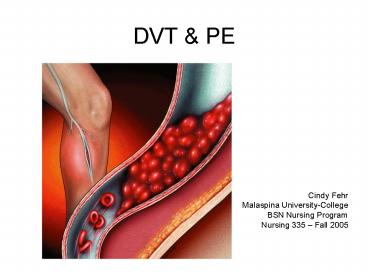
DVT & PE Cindy Fehr Malaspina University-College BSN Nursing Program Nursing 335 Fall 2005 FACTS Orthopedic injuries place at risk DVT formation of thrombus ... – PowerPoint PPT presentation
Number of Views:425
Avg rating:3.0/5.0
Title: DVT
1
DVT PE
- Cindy Fehr
- Malaspina University-College
- BSN Nursing Program
- Nursing 335 Fall 2005
2
FACTS
- Orthopedic injuries place at ? risk
- DVT formation of thrombus in deep vein,
typically in lower extremity - PE thrombus that travels into the pulmonary
circulation
3
Pathophysiology of DVT
- Prevention is key
- Identify those at high risk
- Virchows triad ? changes in blood coagulability,
changes in vessel wall, changes in blood flow - Prophylactic tx with oral anticoagulant or LMW
heparin - Intermittent pneumatic compression devices (calf
compressors) or elastic stockings (TEDS) - Early ambulation
- Early identification treatment of DVTs
- treat with vena cava filters
4
Coagulation Pathways
- Extrinsic Pathway - activated when tissue trauma
occurs - Intrinsic Pathway - activated under conditions
such as stress, anxiety, or fear, and in the
absence of external tissue injury - stage one cascade - either the intrinsic or
extrinsic pathway ends with the formation of
prothrombin-converting factor - stage two prothrombin-converting factor begins
series of chemical interactions ? slowly converts
prothrombin to thrombin. - Stage three fibrinogen interacts with thrombin to
form fibrin. - erythrocytes, phagocytes, and microorganisms,
also collect at the site to complete thrombosis
development
5
predisposing factors of DVT
- advanced age
- trauma
- spinal cord injury
- immobilization
- myocardial infarction
- heart failure
- stroke
- previous thromboembolic disease
- thrombotic abnormalities
- obesity
- pregnancy
- constricted clothing
- homocystinuria
- systemic lupus erythematosus
- inflammatory bowel disease
- central venous catheter use
- oral estrogen use
6
Pulmonary Embolism
- About 10 of PEs result in immediate death
- a futher 20 kill later if they're untreated
- Once treated, the mortality rate drops to 3.
7
Source Nucleus Medical Art Online
8
Venous thromboembolism (PE)
- a serious and commonproblem that can and should
be prevented - A serious problem
- 80 of PE occur without signs
- 2/3 of deaths occur within 30 minutes
- A common problem
- One in 100 hospitalized patients dies of PE
- Can should be prevented
9
These pulmonary emboli removed at autopsy look
like casts of the deep veins of the leg where
they originated. Source www.DVT.ORG
10
What increases the chances of having a pulmonary
embolism?
- Older adults, especially those who are bedridden
- People who have or have had cancer
- Anyone who has recently undergone surgery,
especially in the abdomen - Family history of pulmonary embolism
- Obesity
- Recent fracture of the pelvis or legs
- Pregnant women and women who have recently given
birth - Oral contraceptive use
11
The main symptoms of a Pulmonary Embolus are
- Chest pain
- Typically under the breastbone or to one side
- Typically a sharp stabbing pain
- Typically made worse when breathing in
- Shortness of breath
- A mild temperature (typically 38C), with
sweating - Rapid pulse
- A dry cough, sometimes with blood (usually small
amounts, sometimes more) - A feeling of anxiety
12
There are three main tests which can be
performed
- D-dimer represents fibrin split products that
are released into the blood stream when there are
clots in the blood stream - Chest X-ray helpful to eliminate other
possibilities, but 25 of PEs don't show up on an
ordinary x-ray - Ventilation Perfusion scan (V/Q scan) good
initial screening test, but notoriously difficult
to analyse - CT scan the definitive diagnostic tool
Source American Family Physician Journal Sept.
2001
13
Treatment
- to thin the blood using anti-coagulants.
- initial injection (bolus) of heparin, followed by
a heparin infusion for several days - although heparin provides immediate protection,
it has an extremely low half-life, so it only
protects you while you're being infused with it - a longer-lasting anti-coagulant is used for
ongoing protection. (warfarin) - on heparin until blood tests (INR test) confirms
that warfarin is keeping your blood two or three
times thinner than usual
14
Treatment cont.
- Less common treatments include
- These options are used when anti-coagulation is
proving ineffective or where the clot is so
severe that it is too dangerous to wait for the
clots to dissolve in the thinned blood - clot-filters
- clot-busting drugs
- surgery
15
Prevention
- To help prevent development of blood clots in the
venous system - Pressure stockings
- early ambulation
- low dose heparin
- use of sequential compression


