BIOL316 Lectures 20 - PowerPoint PPT Presentation
1 / 38
Title: BIOL316 Lectures 20
1
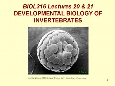
BIOL316 Lectures 20 21 DEVELOPMENTAL BIOLOGY OF
INVERTEBRATES
(Gould and Keeton, 1996. Biological Sciences, Vol
1, Norton, New York and London)
2
Biol316 Lecture 20 Invertebrate Development Part 1
development
- is the process by which fertilised eggs are
transformed into functioning multicellular
organisms - is driven by cell division, cell migration and
cellular differentiation - is controlled by the activation of pre-defined
cellular programs in response to precise
positioning signals
3
- EVO-DEVO
- the major evolutionary events that gave rise to
the different invertebrate phyla can be traced to
the evolution of developmental processes - therefore, the phylogenetic diversification of
invertebrates is reflected by the progressive
diversification of their developmental processes - Haeckel ontogeny recapitulates phylogeny
Fig 18.2 Ernst Haeckel (1834-1919)
Fig 18.1 Haeckels phylotypes
http//www.angelfire.com/mi/dinosaurs/ontogeny.htm
l
http//www.angelfire.com/mi/dinosaurs/ontogeny.htm
l
4
http//en.wikipedia.org/wiki/Ernst_Haeckel
5
the triploblast bauplan
- most invertebrates are triploblastic they have
3 primary germ layers endoderm, ectoderm and
mesoderm - current phyla arose from a common ancestor
- that ancestor is depicted by the triploblast
bauplan, which is roughly equivalent to a stem
platyhelminth
6
- here, I describe key developmental processes
- most often I refer to the 2 best studied
invertebrate models of development sea urchins
(Strongylocentrotus purpuratus Heliocidaria
erythrogramma),which are deuterostomes, and the
vinegar fly, Drosophila melanogaster - an
arthropod
Fig 18.3 A molecular phylogeny of the animal
kingdom, http//www.bio.miami.edu/dana/106/106F03_
11.html
7
5 key developmental processes
- Fertilization
- Cleavage
- Gastruation/neurulation
- Patterning/body plan
- formation
- 5. Tissue differentiation
lecture 20
lecture 21
- the nature and complexity of these processes is
dependent on the phylogenetic position of an
animal
8
1. Fertilization in water
- sperm/egg interactions are critical,
particularly among marine broad spawners, in
which sperm have to find eggs of their own
species in the big bad ocean - Broad spawning interactions
- are regulated by
- synchronised spawning
- based on temperature, moon,
- day length etc
- chemical chemoattraction e.g. L-tryptophan in
abalone - species specific molecular interactions at
sperm/egg surfaces e.g. lysin and VERL in abalone
Fig 18.4 Fertilisation, Gould and Keeton, 1996.
Biological Sciences, Vol 1, Norton, New York and
London.
9
1. Fertilization on land
Fig 18.5 Spermatophore transfer in scorpions
http//www.beepworld3.de/members22/scorpionida/22
.htm
- sperm needs to be protected from desiccation
- spermatophore (tough casing)
- also used by aquatic animals (cephalopods,
decapods) - transfer into female genital tract
- sperm in seminal fluid
- Some animals deposit spermatophore on substrate
and females pick it up - Eg. Scorpions colembola
10
1. Fertilization on land
- Sperm transfer
- Direct if intromittant organ connected to testes
- eg. penis (aedeagus)
- Indirect if intromittant organ not connected to
testes - eg. spider pedipalps,
Fig 18.6 Spider pedipalps http//images.opentopia
.com/enc/257/256447/20040817_010343_DSC5954.jpg
11
2. Cleavage
- rapid mitotic division immediately after
fertilisation - division is rapid because G1 and G2 stages of
the cell cycle are reduced to the point where
cells do not have a chance to increase in volume
after division - therefore, multicell embryos are often no bigger
than an unfertilised egg
Fig. 18.7 Cleavage in the purple sea urchin, S.
purpuratus (http//worms.zoology.wisc.edu/urchins)
12
- cleavage can occur in two orientations
Fig. 18.8a Spiral cleavage in protostomes,
http\MyFile\Courses\Biology\Bio211\notes for
website\04\04 EMBRYOLOGY
cleavage plane rotates on a spiral axis between
divisions so, the cells of the upper layer are
located in the angles between the cells of the
lower layer
Fig. 18.9b Radial cleavage in deuterostomes http\
MyFile\Courses\Biology\Bio211\notes for
website\04\04 EMBRYOLOGY
cleavage plane alternates at 90 angles between
divisions - so the cells of the upper layer are
located directly above the cells of the lower
layer
13
- cleavage patterns are affected by the amount of
yolk in an egg
e.g. sea urchins
Fig. 18.10
e.g. insects
http\MyFile\Courses\Biology\Bio211\notes for
website\04\04 EMBRYOLOGY
Cleavage in Drosophila
14
- the generalised fate of cells is determined
during cleavage
epidermal (Epi), neuron (Neu), structural (Str),
and death (X)
Fig. 18.11 Cell lineage of the nematode C.
elegans, http//scienceblogs.com/pharyngula/2006/0
3/modeling_metazoan_cell_lineage.php
15
Fig 18.12 A cleavage fate map for a polychaete
worm, Arenicola marina, Neilsen, C. 2004. Journal
of Experimental Zoology. 302B 35-68
16
- the fate of cells can be either fixed
(determinant) or plastic (indeterminant) - deuterostome embryonic stem cells remain
totipotent long into development, whilst the fate
of protostome stem cells is fixed very early in
cleavage and cannot be reversed
Fig 18.13 Fate determination in protostome and
deuterostomes, http\MyFile\Courses\Biology\Bio211
\notes for website\04\04 EMBRYOLOGY ,
deuterostome
protostome
17
- the end product of cleavage is a spherical
blastula with a fluid filled interior (blastocoel)
Fig 18.14a. Sea urchin (S. purpuratus) blastula
(http//worms.zoology.wisc.edu/urchins/SUgast_intr
o.html)
Fig 18.14b. Drosophila blastula
(http//flybase.net/images/lk/Embryogenesis/Gastru
lation/Gastrulation-Lateral/GLV-1-lbl.jpeg)
18
3. Gastrulation
- the first series of cellular migrations in an
embryo - forms the gut and primary germ layers
- provides embryos with an anterior/posterior axis
- is the first stage of patterning or body plan
formation
Fig 18.15 Gastrulation in S. purpuratus
(http//worms.zoology.wisc.edu/urchins/SUgast_move
ments1.html)
19
- gastrulation results from the migration of cells
from the surface of the blastula into the
blastocoel - there are a number of ways in which this
migration can occur, determined largely by the
residual yolkiness of the embryo
Fig 18.16 Three different methods of
gastrulation, http//worms.zoology.wisc.edu/urchin
s/SUgast_intro.html
20
- sea urchins have little yolk and so use
invagination - invagination is preceded by the detachment of
primary mesenchyme cells (PMCs) into the
blastocoel these cells will form part of the
mesoderm
PMCs
Fig. 18.17 The beginnings of invagination in S.
purpuratus, http//worms.zoology.wisc.edu/urchins/
SUgast_primary.html
21
- after PMC migration, a group of cells at the
vegetal pole of the blastula begin to bend inward
forming the archenteron (presumptive gut) and the
blastopore (entrance to archenteron - bending is caused by the contraction of actin
microfilaments at the apical surface of the cells
B
actin microfilaments
A
C
http//worms.zoology.wisc.edu/urchins/su_veg_plate
_SEM_72dpi
D
http//worms.zoology.wisc.edu/urchins/SUgast_ingre
ssion.html
Fig 18.18 Invagination
http//worms.zoology.wisc.edu/urchins/su_veg_plate
_phall_72dpi
Fig 13.5. Gould and Keeton, 1996. Biological
Sciences, Vol 1, Norton, New York and London.
22
- the archenteron continues to push through the
blastocoel until it reached the opposite side of
the blastula where it fuses with the wall of the
blastula - this forms a flow through gut and leaves the
embryo with 3 primary germ layers of cells
mesoderm
http//worms.zoology.wisc.edu/urchins/SUgast_ingre
ssion.html
http//worms.zoology.wisc.edu/urchins/SUgast_ingre
ssion.html
ectoderm
Fig 18.19 The archenteron
Fig 18.20 The three primary germ layers
endoderm
gut
23
- the order in which the mouth and anus are formed
during gastrulation differs between protostomes
and deuterostomes, hence their names
Protostome mouth first Deuterostome mouth
second
Fig 18.21 Mouth and anus formation in protostomes
vs deuterostomes, http\MyFile\Courses\Biology\Bio
211\notes for website\04\04 EMBRYOLOGY
24
Gastrulation
25
Biol316 Lecture 21 Invertebrate Development Part 2
4. Patterning
- processes that begin with gastrulation establish
the basic body plan, or pattern, for the final
individual - this pattern provides the embryo with a 3
dimensional image of itself that is used to guide
the final development of organs, building limbs,
wings, eyes etc (i.e. tissue differentiation) - most often, the body plan is based around the
anterior/posterior, dorso-ventral axis formed by
the gut and nervous system - these axes allow the body to be broken up into
distinct domains, each of which goes on to
develop relatively independently
26
morphogens and segmentation in Drosophila
- like many invertebrates (all arthropods,
annelids, urochordates), in Drosophila the body
plan is based on segmentation - in Drosophila each segment (6 head, 3 thorax and
8 abdomen) develops specific functions in the
adult - during development those segments are
established by the release of morphogens
proteins that establish concentration gradients
throughout the embryo to provide cells with
precise 3-dimensional positioning information
27
- segmentation begins with the deposition of mRNAs
for the morphogens bicoid and nanos into the egg
before fertilisation. - these mRNAs are translated into their proteins
after fertilisation to produce - an anterior to posterior concentration gradient
of the morphogen, bicoid - a posterior to anterior concentration gradient of
the morphogen, nanos
Fig 20.1 Primary morphogens in Drosophila, Gould
and Keeton, 1996, Biological Sciences, Norton,
New York and London.
nanos
bicoid
concentration of morphogen
posterior
anterior
28
- bicoid and nanos have opposite effects on a gene
called hunchback - bicoid is a transcription factor that activates
hunchback - nanos inhibits the translation hunchback mRNAs
- these effects combine to produce a high levels
of hunchback protein at the anterior of the
embryo and low levels toward the posterior
Fig. 20.2 The effect of bicoid and nanos on
hunchback expression, red arrow activation,
blue bar inhibition, file///C/WORK/UNDERGRAD/B
IOL20316/Segmentation.html
29
-
- the combined concentration gradients of bicoid,
nanos and hunchback sequentially activate or
inhibit other morphogen genes in increasingly
sharply-defined regions of the embryo - the final complex language of morphogens
precisely defines the 3 dimensional structure of
an embryo allowing cells to determine precisely
which segment, and which area of that segment,
they are in
embryo stained for eve expression
Fig 20.3 The gene, eve, is expressed in 7
segments in the abdomen. The morphogens bicoid
(bcd) and hunchback (hb) stimulate the
transcription of eve, whilst giant (gt) and
Krüppel (Kr) inhibit eve expression,
http/www.WORK/UNDERGRAD/BIOL20316/Segmentation.h
tml
30
5. Tissue differentiation - homeotic genes
- the genes that are ultimately activated by the
positioning signals provided by morphogens are a
group of about 20 segment-specific homeotic
genes - all of these homeotic genes share homeobox
domains, which allow them to act as transcription
factors
Fig 20.4 Amino acid sequence for typical homeotic
gene, Antennapedia, showing its homeobox domains
in orange, http//www.WORK/UNDERGRAD/BIOL20316/Ho
meoboxGenes.html
31
- most homeotic genes are organised in two tight
chromosomal clusters, the Antennapedia and
Bithorax clusters - the segment in which they are expressed depends
on the order that they appear in the cluster
Fig 20.5. Sites of expression for members of the
Antennapedia cluster of homeotic genes,
http//www/WORK/UNDERGRAD/BIOL20316/Segmentation.
html
32
- homeotic genes act as selectors they
activate the construction of segment-specific
traits like wings and legs on the thorax,
antennae and eyes on the head etc - for example, antennapedia (Antp) activates the
construction of legs on the thoracic segments - Antp is expressed in the thorax, but turned off
(repressed) in the head
Fig 20.6 Flies with the antennapedia mutation
express Antp in head segments, Gould and Keeton,
1996, Biological Scienes, Norton, New York and
London.
33
Larvae and metamorphosis
- in many invertebrates, development is further
complicated by the presence of intermediate
larval stages between egg and adult - larval stages have significant benefits, like
dispersal in species that have sessile adults - but they require one additional step in
development metamorphosis into the final adult
form
B
Fig 20.7 Invertebrate larvae A. insect. B.
echinoderm pluteus (Gould and Keeton, 1996,
Biological Scienes, Norton, NewYork and London
http//www.microscopy-uk.org.uk/mag/indexmag.html?
http//www.microscopy-uk.org.uk/mag/artjul00/urchi
n1.html)
A
34
- the complexity of metamorphosis depends on the
relationship between larvae and adults
ametabolous
hemimetabolous
holometabolous
Fig 20.8 3 different generalised life history
strategies in insects, http//www.ndsu.nodak.edu/e
ntomology/topics/growth.htm
35
- in some cases, metamorphosis can be as simple as
adding new, almost identical segments to an
elongating body
Fig 20.9 Metamorphosis of a polychaete
trochophore larva, Neilsen, C. 2004. Journal of
Experimental Zoology. 302B 35-68
36
- however, holometabolous metamorphosis requires
elaborate systems to construct adults - in insects like Drosophila, selector genes that
control adults secondary characteristics form
imaginal discs in larvae as repositories of adult
characteristics
Fig 20.10 The construction imaginal discs in
Drosophila larvae is controlled by selector
(homeotic) genes, Gould and Keeton, 1996,
Biological Sciences, Norton, New York London.
37
- important things that Im NOT going to talk
about - the formation of body cavities (coelom and
pseudocoelom) - neurolation (formation of the nervous system)
- development of buds in asexually reproducing
species - development of zooids in colonial organisms
38
links
Development in general http//www.personal.psu.edu
/faculty/w/x/wxm15/Online/Zoology20Unit/zoology_l
inks.htm Sea urchin development http//worms.zool
ogy.wisc.edu/urchins/SUIntro.html http//www.stanf
ord.edu/group/Urchin/ani-plus.htm Drosophila
development http//flybase.net/allied-data/lk/inte
ractive-fly/aimain/1aahome.htm Homeoboxes
http//users.rcn.com/jkimball.ma.ultranet/Biology
Pages/H/HomeoboxGenes.html Metamorphosis http//
www.ndsu.nodak.edu/entomology/topics/growth.htm


