Alexandre G' de Brevern - PowerPoint PPT Presentation
1 / 38
Title:
Alexandre G' de Brevern
Description:
INSERM UMR-S 726 - quipe de Bioinformatique G nomique et Mol culaire (EBGM) ... a-helix core : aliphatic residues (Leucine, Valine and Isoleucine) aromatic ... – PowerPoint PPT presentation
Number of Views:127
Avg rating:3.0/5.0
Title: Alexandre G' de Brevern
1
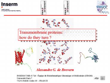
Transmembrane proteins how do they turn ?
- Alexandre G. de Brevern
INSERM UMR-S 726 - Équipe de Bioinformatique
Génomique et Moléculaire (EBGM) Université Paris
7 75251 PARIS Cedex 05 FRANCE
2
Transmembrane proteins
- 1- Introduction
- 25 of protein sequences
- Essential biological function
- Target of 70 patent medecine (drugs)
- 1 of the protein structures of the Protein
- DataBank
3
Transmembrane proteins
2- Specific topology
4
Transmembrane proteins
3 The simple question
Jean Pylouster (Master 2nd year - Bioinformatics)
5
Transmembrane proteins
3 The simple question
Is it so simple ?
At the beginning
Why this question ?
6
Transmembrane proteins
4 Protein structures
7
Secondary structures
H bonds
Dihedral angles
a helix
b sheet
distance
angles
3D structure
Dihedral angles
Protein
Volume
8
Secondary structures
- Different assignment methods
- Greer Levitt (1977) Distances
- DSSP (Kabsch Sander, 1983). Hydrogen bonds
- DEFINE (Kundrot Ridchards, 1988).
Distances - PCURVE (Sklenar, Etchebest and Lavery, 1989).
Axes - CONCENSUS (Colloch, Etchebest et al., 1993).
Mean values of 3 previous methods - STRIDE (Frishmann Argos, 1995).
Hydrogen bonds / dihedral angles - PSEA (Labesse et al., 1997).
Distances / angles - PROSS (Srinivasan Rose, 1999).
Dihedral angles - XTLSSTR (King Johnson , 1999).
Distances / angles - DSSPcont (Andersen et al., 2001). Hydrogen bonds
/ dihedral angles - SECSTR (Fodje Al-Karadaghi, 2002). Hydrogen
bonds / dihedral angles - VORO3D (Dupuis et al., 2004).
Volume - KAKSI (Martin et al., 2005).
Distances / dihedral angles - SEGNO (Cubellis et al., 2005).
Angles / multiple - Beta-Spider (2005), PALSSE (2005), Delaunay
tessalation (2005)
9
Les structures secondaires
No simple consensus between methods
assignment of Hhai Méthyltransferase (code PDB)
10MH with DSSP, STRIDE, PSEA, DEFINE, PCURVE,
XTLSSTR and SECSTR.
L. Fourrier, C. Benros A.G. de Brevern (BMC
Bioinfo, 2004)
10
Les structures secondaires
J. Martin, G. Letellier, J.F. Taly, A. Martin,
A.G. de Brevern J.F. Gibrat (BMC Structural
Biology, 2005)
http//migale.jouy.inra.fr/mig/mig_fr/servlog/kaks
i/
11
Transmembrane proteins
- PDBTMProtein Data Bank of Transmembrane Proteins
http//pdbtm.enzim.hu/ - Tusnády GE, Dosztányi Zs and Simon I (2004)
Transmembrane proteins in protein data bank
identification and classification. Bioinformatics
20, 2964-2972.
6 helices !!
12
Transmembrane proteins
- Orientations of Proteins in Membranes (OPM)
database - http//opm.phar.umich.edu/
- Lomize AL, Pogozheva ID, Lomize MA, Mosberg HI
(2006) Positioning of proteins in membranes A
computational approach. Protein Science 15,
1318-1333. .
13
Transmembrane proteins
5 The simple simple principle
Non-redundant structural Databank
14
Transmembrane proteins
6 The beautiful databank
Zhou Zhou (Protein Sci., 2003) 73 protein
structures
15
Transmembrane proteins
6 The beautiful databank
- Elimination of 1/4th of the proteins
- -NMR structures 10
- - Ca structures 2,
- Transmembrane region(s) not present 4
- no PDB or sequence found 1
1FDM
putative position of helix
16
Transmembrane proteins
7 Comparison of secondary structure assignment
3D structure of the bacteriorhodopsin assigned by
different SSAMs. (a) DSSP, (b) STRIDE, (c)
SECSTR, (d) SEGNO, (e) KAKSI, (f) ZZ, (g) PSEA,
(h) XTLSSTR, (i) PCURVE and (j) the Protein
Blocks.
17
Transmembrane proteins
7 Comparison of secondary structure assignment
3D structure of the bacteriorhodopsin assigned by
different SSAMs
18
Transmembrane proteins
7 Comparison of secondary structure assignment
3D structure of the bacteriorhodopsin assigned by
different SSAMs, is also shown the prediction
done by Zhou and Zhou
19
Transmembrane proteins
7 Comparison of secondary structure assignment
AA WDKYAQEVYEMNFGEKPEGDITQV DSSP
CCCHHHHHHHHHHHCCCCCCCCCC STRIDE
CCHHHHHHHHHHHHHHCCCCCCCC PSEA
CCHHHHHHHHCCCCCCCCCCCCCC PCURVE
CCHHHHHHHHHHCCCCCCCCCCCC
DSSP CCCHHHHHHHHHHHCCCCCCCCCC STRIDE
CCHHHHHHHHHHHHHHCCCCCCCC
20
Transmembrane proteins
7 Comparison of secondary structure assignment
21
Transmembrane proteins
- 7 Comparison of secondary structure assignment
- Classical distribution in regards to SSAMs
observed for globular proteins. - Protein Blocks and ZZ assignment slightly
different - Surprisingly DSSP done the mean shortest helices
(in fact due to numerous kinks in the structure,
found also with other SSAMs). - 8 of linear helices, 50 of curved and 29 of
kinked.
22
Transmembrane proteins
8 Sequence structure relationship Analysis
of amino acid over- and under-representation
23
Transmembrane proteins
8 Sequence structure relationship a-helix
N cap
Coil helix ND(G) PW EW
24
Transmembrane proteins
8 Sequence structure relationship a-helix
Helix () ILFW (-) DEKP
25
Transmembrane proteins
8 Sequence structure relationship a-helix
C cap
helix Coil - NG P P
26
Transmembrane proteins
- 8 Sequence structure relationship
- a-helix N cap ND(G)S0 by PW1 and EW2
- a-helix core aliphatic residues (Leucine,
Valine and Isoleucine) aromatic
residues (Tryptophan and Phenylalanine)
hydrophobic. - a-helix C cap NG1 P2 P3
- So classical results. Some limited shifts. No new
patterns. - ZZ strongest informativity for core, lowest for
capping regions.
27
Transmembrane proteins
9 Prediction methods Principle
X 1000 independent simulations
15 aa
Matrix H
Matrix C
28
Transmembrane proteins
9 Prediction methods
29
Transmembrane proteins
9 Prediction methods Thus, automatic secondary
structure final prediction rates using only
single sequence are within a range from 78.26 to
80.95. A structural alphabet approach gives a
slight better prediction (Qtot equals to
81.46), while secondary structure assignment
used for benchmark set, i.e. ZZ, gives a
prediction rate of 86.27. This last remark is
striking as it corresponds to a difference of 5
with the best SSAM, i.e. STRIDE, and 6.4 with
DSSP, the most classical SSAM.
30
Transmembrane proteins
9 Prediction methods The behaviour of ZZ is
mainly due to a lower number of helix
residues. In fact, it predicts 10 less helix
than other approaches while its helix frequency
is only 5 lower.
31
Transmembrane proteins
- 10 Conclusion
- SSAMs have similar behaviors for TMb proteins
even if they have been conceived for globular
proteins. - They do not highlight new amino acid patterns.
- Prediction is strongly dependant to assignment
methods. - Always problems to evaluate TMb prediction
quality.
32
Transmembrane proteins
Thank you
33
Transmembrane proteins
Supplementary slides
34
PBs
35
Structural alphabet
A structural alphabet is a set (or library) of
small prototypes which approximate every part of
the protein structures. They are composed by a
limited number of recurrent structural elements
of proteins. The associations between these
structural "letters" are governed by logic rules
and form the words of protein structures. A
structural alphabet has no a priori in regards to
the secondary structures, i.e. it is not a
categorization of the coil state.
de Brevern A.G., Camproux A.C., Hazout S.,
Etchebest C., and Tuffery P. (2001), Protein
structural alphabets beyond the secondary
structure description, Recent Adv. In Prot. Eng.,
1319-331 .
36
Structural alphabet
de Brevern A.G., Etchebest C. Hazout, S.
(2000), Bayesian probabilistic approach for
prediction backbone structures in terms of
protein blocks, Proteins, 41(3)271-287.
37
Structural alphabet
gt153L secondary structuresHHHHCCCCCCCCCCCCCCCHH
HHHHCCCCCCCHHHHHHHHHHHHHHHHHHHHHHHCCCCCCCHHHHHHHH
HCCCCCCCCCCCCCCCCCCCCCCCCCCCCCCCCCHHHHHHHHHHHHHH
HHHHHHHHCCCCCHHHHHHHHHHHCCCCEEEEECCCCCCCCHHHHHHHH
HHHHHHCCCC
gt153L structural alphabet ZZmnopfklpccebjafklmmm
nopabecjklmmmmmmmmmmmmmmmmmmmmmmmmmnopafklmmmmmmm
mnooolapgehiafkopagcjkopafklmccehjfklmklmmmmmmmmm
mmmmmmmmmmmmbcfklmmmmmmmmmmnomklmnbfklmmgoiahilm
mmmmmmmmmmmmmnoZZ
38
Structural alphabet
? local protein structure approximation (Proteins
2000, ISB 2005) ? local structure
prediction (Proteins 2000, 2005, ISB 2004, J
Biosc 2006) ? local protein structure
approximation for longer fragments (Protein
Science 2002, Bioinformatics 2003) ? local
protein structure prediction for longer
fragments (Proteins 2006a) ? Superimposition of
protein structures (Proteins 2006b, Nucleic Acid
Res 2006) ? Molecular modeling (Bioch Biophys
Acta 2005)


