19 The Urinary System - PowerPoint PPT Presentation
1 / 37
Title:
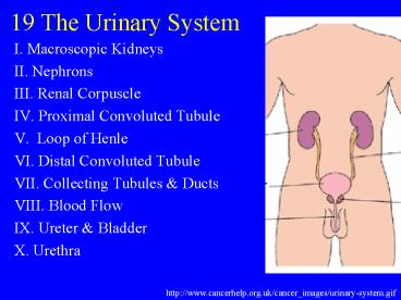
19 The Urinary System
Description:
Secondary processes interdigitate around glomerular capillaries. ... http://images.webmd.com/static54/images/hwstd/medical/urology/n5550380.jpg ... – PowerPoint PPT presentation
Number of Views:78
Avg rating:3.0/5.0
Title: 19 The Urinary System
1
19 The Urinary System
- I. Macroscopic Kidneys
- II. Nephrons
- III. Renal Corpuscle
- IV. Proximal Convoluted Tubule
- V. Loop of Henle
- VI. Distal Convoluted Tubule
- VII. Collecting Tubules Ducts
- VIII. Blood Flow
- IX. Ureter Bladder
- X. Urethra
http//www.cancerhelp.org.uk/cancer_images/urinary
-system.gif
2
I. Macroscopic Kidneys
- A. Capsule hilus (19.1)
- B. Renal sinus
- 1. renal pelvis
- 2. major calyces
- 3. minor calyces
- C. Renal medulla
- 1. renal pyramids
- a. papilla
- 2. Renal column
- D. Renal cortex
3
II. Nephrons
- A. Components (19.1)
- 1. renal corpuscle
- 2. PCT
- 3. loop of Henle
- 4. DCT
- 5. (collecting tubule duct)
4
II. Nephrons
- B. Relationship to cortex and medulla (19.1)
5
III. Renal Corpuscle
- A. Glomerulus (19.3)
- 1. afferent arteriole
- 2. efferent arteriole
- 3. vascular pole
- 4. fenestrated capillaries (19.4)
6
III. Renal Corpuscle
- B. Bowmans capsule
- 1. visceral layer
- a. podocytes (19.3)
7
III. Renal Corpuscle
- B. Bowmans capsule
- 1. visceral layer
- a. podocytes
- 1) primary processes (19.5)
8
III. Renal Corpuscle
- B. Bowmans capsule
- 1. visceral layer
- a. podocytes
- 2) secondary processes (pedicels, foot
processes) (19.5,19.6)
9
III. Renal Corpuscle
- Secondary processes interdigitate around
glomerular capillaries. The narrow space between
processes is the filtration slit. 19-5
10
III. Renal Corpuscle
- B. Bowmans capsule
- 1. visceral layer
- a. podocytes
- 3) filtration slits (19.7)
11
III. Renal Corpuscle
- B. Bowmans capsule
- 1. visceral layer
- a. podocytes
- 3) filtration slits
- a) 25 nm wide (19.8)
- 4) basement membrane
12
III. Renal Corpuscle
- B. Bowmans capsule (19.3)
- 2. urinary space
- 3. parietal layer
- 4. urinary pole
13
III. Renal Corpuscle
- B. Bowmans capsule
- 5. EM view (19.4)
14
III. Renal Corpuscle
- C. Structure/function 19.7
- 1. glomerular filtrate
- 2. most blood components smaller than 70 kDa
15
IV. Proximal Convoluted Tubule
- A. Overview (19.3,19.13)
- 1. begins at urinary pole
- 2. simple cuboidal epithelium
- 3. longer than DCT
16
IV. Proximal Convoluted Tubule
- B. Cell structure
- 1. brush border (19.14,19.15)
- a. microvilli 1 mm
17
IV. Proximal Convoluted Tubule
- B. Cell structure
- 2. basal border (19.4)
- a. membrane invaginations
- 1) Na/K-ATPase
- b. parallel mitochondria
- (recall salivary glands)
18
IV. Proximal Convoluted Tubule
- C. Function 19.16
- 1. pinocytosis
- 2. active ion transport
- 3. Osmosis
- 4. exocytosis
19
IV. Proximal Convoluted Tubule
- C. Function 19.16
- 4. reabsorption
- a. all glucose
- b. amino acids
- c. 85 NaCl H20
- d. PO4, Ca
- e. protein
- 5. secretion/excretion
20
V. Loop of Henle
- A. Components (19.16)
- 1. thick descending limb
- 2. thin descending limb
- 3. thin ascending limb
- 4. thick ascending limb
cortex
medulla
21
V. Loop of Henle
- B. Cell structure
- 1. thick descending PCT
- 2. thick ascending DCT
- 3. thin limbs (19.18,19.19)
22
V. Loop of Henle
- C. Function 19-16
- 1. capability of forming hypertonic urine
- a. water retention
- 2. thin descending limb
- a. permeable to water
- 3. ascending limb
- a. impermeable to water
23
VI. Distal Convoluted Tubule
- A. Location (19.16)
- 1. continuation of thick ascending limb
- 2. cortex
- 3. vascular pole of renal corpuscle
- a. juxtaglomerular region
24
VI. Distal Convoluted Tubule
- B. Cell structure (19.16,19.19)
- 1. simple cuboidal epithelium
- 2. cells smaller than PCT
- a. more nuclei visible/XS
- 3. lack brush border
25
VI. Distal Convoluted Tubule
- B. Cell structure (19.19)
- 4. less acidophilic than PCT
- 5. larger lumen than PCT
- 6. similar basal infoldings
26
VI. Distal Convoluted Tubule
- C. Function 19.16
- 1. under influence of aldosterone
- 2. absorb Na (and H2O)
- 3. secretes K, H, NH4
27
VI. Distal Convoluted Tubule
- D. Juxtaglomerular region
- 1. macula densa of DCT (19.21)
- a. DCT cells columnar
- b. able to sense flow and ionic conc.
- c. influences afferent arteriole
constriction, glomerular filtration, renin
secretion
28
VI. Distal Convoluted Tubule
- D. Juxtaglomerular region
- 2. JG apparatus (19.25)
- a. modified smooth muscle of afferent
arteriole - b. secretory granules
- c. protein synthesizing organelles
- d. synthesize renin
- e. effect increase Na and Cl- absorption
distal tubules
29
VII. Collecting Tubules Ducts
- A. Cell structure
- 1. cuboidal to columnar (19.22)
- 2. weakly staining
- 3. few organelles
30
VII. Collecting Tubules Ducts
- A. Cell structure
- 4. no basal striations (19.23)
- 5. clear intercellular borders
- 6. empty into minor calyx at renal papilla
31
VII. Collecting Tubules Ducts
- B. Function 19-16
- 1. ADH (vasopressin)
- a. causes epithelium to be permeable to H2O
- b. reabsorption of H2O
32
VIII. Blood Flow
- A. Renal artery (19.26)
- B. Interlobar arteries
- C. Arcuate arteries
- D. Interlobular arteries
33
VIII. Blood Flow
- E. Afferent arterioles (19.26)
- F. Efferent arterioles
- G. Peritubular capillary network
- G. Vasa recta
34
IX. Ureter Bladder
- A. Ureter 19-28
- 1. mucosa
- a. transitional epithelium
- b. lamina propria
- 2. muscularis
- a. inner longitudinal
- b. outer circular
- 3. adventitia
35
IX. Ureter Bladder
- B. Urinary bladder
- 1. mucosa (19.27)
- a. transitional epithelium
- b. lamina propria
- 2. muscularis
- a. fibers run in all directions
- b. no layers
- 3. adventitia / serosa
36
X. Urethra
- A. Male urethra (22.1)
- 1. prostatic urethra
- 2. membranous urethra
- 3. pendulous urethra
- a. pseudostratified / columnar epithelium
- b. corpus spongiosum
- 4. bulbous urethra
37
X. Urethra
- B. Female urethra
- 1. pseudostratified columnar, 4-5 cm long
- 2. stratified squamous
http//images.webmd.com/static54/images/hwstd/medi
cal/urology/n5550380.jpg


