Pr - PowerPoint PPT Presentation
Title: Pr
1
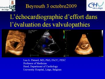
Beyrouth 3 octobre2009
Léchocardiographie deffort dans lévaluation
des valvulopathies
Luc A. Piérard, MD, PhD, FACC, FESC Professor of
Medicine Head, Department of Cardiology University
Hospital, Liège, Belgium
2
Exercise echo in valvular heart disease
- Valvular heart disease is usually considered
static - Management relies upon resting evaluation only
- Most valve disease have a dynamic component
- Exercise testing
- can induce symptoms
- reveals the dynamic of the valve and the
ventricle
3
Functional assessment
- Asymptomatic aortic stenosis
- Mitral stenosis
- Mitral regurgitation
- organic
- ischaemic
- Aortic regurgitation
4
Natural history of aortic stenosis
0 40 50 60 70 80
Age (year)
Symptomatic severe AS indication of AVR (class
I)
5
Risk stratification in asymptomatic AS
- Benign prognosis ?
- Events preceded by symptoms (SD lt 1 /y )
- Symptom or event-free at 2y
- varying 21 to 67
- Higher risk
- age, renal failure, V max gt 4.5 m/s,
inactivity, CAD - rapid increase of Ao velocity gt 0.3m/s/y Ca2
- - 50 of pts deny symptoms
- SD without symptoms, Waiting list ?
- Prognosis if symptomatic at operation ?
Otto Circ 1997, Rosenhek NEJM 2000, Pellika Circ
2005
6
Indications for AVR in AS ACC/AHA Guidelines
2006 ESC 2007
Class Asymptomatic pts with severe AS
and LV systolic dysfunction I (C)
Symptoms or Fall in SBP during Ex IIb (C) (ESC
2007 I C if symptoms IIa C if fall in SBP,
Arrhythmias IIb( C) Likelihood of rapid
progression IIb (C) AVA lt 0.6 cm²
IIb (C) Prevention of sudden death
III
7
Exercise testing in aortic stenosis who?
- - Contra-indicated in symptomatic patients
- In asymptomatic patients
- Class IIb (ACC/AHA)
- Useful (ESC)
8
Risk stratification with exercise testing why?
30 Asymptomatic Pts, age 6214 years, AVA
0.70.2 cm², Peak Grad 7921 mm Hg
POSITIVE TEST Dyspnea,angina,syncope or
near-syncope Fall in SBP during Ex. or rise
lt 20 mmHg Complex ventricular arrhythmias
(VT) gt 2 mm ? ST in comparison to
baseline WG on VHD of the ESC , EHJ 2003
Alborino D et al J Heart Valve D 2002 11204
9
Risk stratification with exercise testing why?
- ? SBP or ? ST
- Limited value
Limiting symptoms during test 46 of 125 pts (37
) No events in pts with AVA gt 1 cm²
Das et al Eur Heart J 2005 261309-13
10
Risk stratification with exercise testing why?
PV 79 if age lt 70 y PV 57 whole
population PV 41 if SAS II Best predictor
Dizziness
Limiting symptoms during test 46 of 125 pts (37
) No events in pts with AVA gt 1 cm²
Das et al Eur Heart J 2005 261309-13
11
Stress echo how?
After exercise During exercise
Dobutamine echo
12
Case 1 75 year-old man, asymptomatic
moderately active AVA 0.65 cm²
HR SBP (bpm) (mmHg) Rest
94 133 Exer 137 154
13
Case 1
No symptoms
? ST Segment
14
Exercise
Rest
E/E 11
E/E 16
PPG 79 MPG 48
PPG 119 MPG 89
AVA 0.65 cm²
AVA 0.59 cm²
PSv 5.1
PSv 7.9
EF 64
EF 71
Increase in mean pressure gradient 41 mmHg
15
Exercise echo in asymptomatic aortic stenosis
Lancellotti, Piérard Circulation 2005112I 377-I
382
16
- Asymptomatic 83-year old man
- Regular follow-up for severe AS
Case 2
Biphasic response
Lancellotti, Piérard Archives of CV diseases 2009
17
Global strain -17.5
Case 3
REST
EF 69.9
PSv 3.8 cm/s
EXERCISE
EF 70.7
PSv 3.7 cm/s
Global strain - 15.5
Impaired contractile reserve
18
Case 4
REST
EXERCISE
M 69 y, AVA 0.7 cm²
Rest Exercise SBP (mmHg) 166 231 HR
(bpm) 85 134 E/E 13.5 31 MPG
(mmHg) 60 68 PSv (cm/s) 4.6 5.1
EF 73
EF 61
Diastolic dysfunction Increase in LV filling P
19
Parameters during exercise Doppler echo
Symptoms Exercise-induced changes mean P
gradient AVA LV EF LV filling P functional
MR pulmonary P Contractile reserve TDI 2D
strain Inducible ischaemia
E/E 12
E/E 35
20
Functional assessment
- Aortic stenosis
- Asymptomatic
- Low-gradient
- Mitral stenosis
- Mitral regurgitation
- Organic
- Ischaemic
- Aortic regurgitation
21
Dobutamine echo in low-gradient AS
Gradient
Area
Fixed true AS Pseudo severe AS Absence of
contractile reserve
(gt 0.3 cm²)
?
22
Rest
EF 23
MPG 23 mmHg PPG 45 mmHg
LVOT 12.1 cm
AVA 0.66 cm²
DOBU 20 µg/kg/min
MPG 44 mmHg PPG 103 mmHg
LVOT 17.4 cm
EF 40
AVA 0.67 cm²
Piérard, Lancellotti Heart 2007
23
Group I contractile reserve Group II no
contractile reserve
Monin et al Circulation 2003108319-24
24
Functional assessment
- Asymptomatic Aortic stenosis
- Mitral stenosis
- Mitral regurgitation
- Organic
- Ischaemic
- Aortic regurgitation
25
Indications of valve repair or replacement
- Severe exercise limitation in pts who deny
symptoms (I) - Haemodynamic changes (IIb if symptoms MVA gt 1.5
cm² ) - (I if asymptomatic MVA 1.5 cm²)
- mean P gradient gt 15 mmHg at exercise (ACC/AHA)
- pulmonary systolic arterial pressure gt 60 mmHg
- Significant increase in MR during test
26
MPG 7.6 MM HG
TTPG 87 MM HG
EXERCISE
27
Functional assessment
- Asymptomatic Aortic stenosis
- Mitral stenosis
- Mitral regurgitation
- Organic
- Ischaemic
- Aortic regurgitation
28
Indications of surgery in asymptomatic MR
- LV ejection fraction lt 60
- LV end-systolic diameter lt 40 mm (ACC/AHA)
- lt 45 mm (ESC)
- Likelihood of valve repair gt 90
29
Organic MR Indications of exercise echo
Exercise Doppler echocardiography is reasonable
(Class IIa) in asymptomatic patients with severe
MR to assess exercise tolerance and the effects
of exercise on pulmonary artery pressure (gt60
mmHg) and MR severity (?) ACC/AHA
2006 Level of Evidence C Not included
in the ESC guidelines 2007
30
LV EDV 250 ml LV ESV 50 ml LV ejection
fraction 80 Regurgitation fraction
56 Forward ejection fraction 24
31
MR may be dynamic
Rest ERO 32 mm² Exercise ERO 52 mm²
32
Degenerative MR
Dynamic MR Exercise-induced pulmonary
hypertension
Exercise PASP gt 60 mmHg is frequent
33
Severe organic MR
Surgery symptoms or EF 60 or ESD 40mm
(ACC/AHA) 45 mm (ESC) or gt 90
likelihood of repair (IIa ACC) (IIb ESC)
LATENT LV DYSFUNCTION
EXERCISE ECHO
34
Predictive value of post-exercise echo
Predictors of post-operative LVEF lt 50
End-systolic volume index post exercise LV
ejection fraction post exercise Exercise-induced
change in LVEF (lt 4)
Leung J Am Coll Cardiol 96281198-205
35
Organic MR latent LV dysfunction ?
Exercise moderate dyspnea
36
Pre-op predictors of post-op EFlt50
Parameter Cut-off AUC Sens Spec Rest LA
volume 78 ml 0.79 64 87 LV ejection
fraction 67 0.48 92 29 Long. Strain
18.1 0.69 77 76 Exercise LV ejection
fraction 70 0.72 69 70 Long. Strain
18.5 0.82 85 76 Ex-induced changes LV
ejection fraction 6.6 0.74 92
53 Long. Strain 1.9 0.80 92 74
Lancellotti, Piérard JASE, 2008
37
Functional assessment
- Asymptomatic Aortic stenosis
- Mitral stenosis
- Mitral regurgitation
- Organic
- Ischaemic
- Aortic regurgitation
38
Ischaemic Mitral Regurgitation
39
Ischaemic MR tethering force
40
The Closing Force
41
Ischaemic MR is dynamic
Rest Increase
at exercise Rest Decrease at exercise
42
- Case History
- 57 y old man
- Patients history
- Posterior AMI treated by thrombolysis PCI
of RCA - NYHA class II
- ACE-I, ß blocker, aspirin, clopidogrel, statin
43
Akinesis of the posterior wall
44
Ischaemic mitral regurgitation
45
VC width 5 mm
6.28 x (0.78)²x 33
ERO
5.8
ERO 22 mm²
RV 22 x 1.85 40 ml
TTPG 36 mmHg
46
No contractile reserve No evidence of inducible
ischaemia
REST
EXERCISE
47
(No Transcript)
48
VC width 7 mm
6.28 x (1.1)²x 33
ERO
6.6
ERO 38 mm²
RV 38 x 1.81 69 ml
TTPG 77 mmHg
49
- Case History
- 57 y old man
- Patients history
- Inferior AMI treated by thrombolysis PCI of
RCA - NYHA class II
- ACE-I, ß blocker, aspirin, clopidogrel, statin
- Clinical evolution
- Acute pulmonary oedema
- furosemide, spironolactone and SL nitrate
- No indication of revascularization
- No current recommendation of mitral
annuloplasty
50
Quantitation
- Doppler method
- PISA method
ERO (mm²) Reg vol (ml)
51
Which quantitative measurement ?
Lebrun, Lancellotti, Piérard JACC 2001 381685-92
52
No relation between MR at rest and
exercise-induced changes
Lancellotti, Lebrun, Piérard JACC 2003, 42,1921
53
Exercise-induced changes in tethering force
Lancellotti, Lebrun, Piérard JACC 2003,
42,1921-28 Giga et al Eur Heart J 2005261860-65
54
Exercise-induced changes in closing force
Lancellotti et al Am J Cardiol 2005961304-7 Laff
itte et al J Am Coll Cardiol 2006472253-9 Piérar
d, Lancellotti Eur Heart J 200627638-40
55
Exercise-induced symptoms
103 fatigue 58 dyspnea
Piérard, Lancellotti N Engl J Med 2006354871-2
56
Dynamic IMR and acute pulmonary oedema
Recent episode of acute pulmonary oedema
Piérard, Lancellotti N Engl J Med 2004
57
Prognosis of ischaemic MR
RR p
ERO lt 20 mm² 1.65 0.049 ERO gt 20 mm²
2.23 0.003
Grigioni Circulation 2001, 103 1759
58
Long-term prognosis of dynamic ischaemic MR
Survival
Decompensated HF
Lancellotti et al Circulation 20031081713-7 Lanc
ellotti, Gérard, Piérard Eur Heart J
2005261528-32
59
Management of ischaemic MR
Clinical HF symptoms, episodes of
decompensation Echo ERO gt 20 mm², Dynamic MR
(ERO gt 13 mm² at exercise) LV remodeling and
sphericity, LV dyssynchrony
Viability /- ischaemia (stress echo or MRI)
TETHERING FORCE
CLOSING FORCE
60
Management of ischaemic MR
Medical CRT PCI CABG Surgical MV
repair Percutaneous annuloplasty
61
Indications of exercise echo in ischaemic MR
- Systolic LV dysfunction and unexplained
- dyspnea
- acute pulmonary oedema
- Risk stratification
- Before bypass grafting in pts with moderate MR
Piérard, Lancellotti Heart 2007
62
Functional assessment
- Asymptomatic Aortic stenosis
- Mitral stenosis
- Mitral regurgitation
- Organic
- Ischaemic
- Aortic regurgitation
63
Test the LV not the valve
Asymptomatic AR
Acute HF
F-UP
REST
Surgery
Post-OP
EXERCISE
AVR if abnormal hemodynamic responses to exercise
(ACC/AHA IIb)
64
Summary
- Beyond symptoms, BP changes and tolerance
- Changes in mean P grad (AS and MS)
- Exercise-changes in MR (mechanisms)
- Changes in LV EF, Contractile Reserve (TDI)
- Myocardial ischaemia
- Changes in LV filling pressure
- Exercise-induced pulmonary hypertension
Valve
Ventricle
Haemodynamic
65
Conclusion
Exercise echocardiography allows a comprehensive
functional evaluation of the dynamic component
and the haemodynamic repercussions of valve
disease


