
Normal Cartilage - PowerPoint PPT Presentation
1 / 6
Title:
Normal Cartilage
Description:
... micrograph of human articular cartilage from the medial ... Chondrocyte clusters (arrows) are visible in the fibrillated. superficial layer of OA cartilage ... – PowerPoint PPT presentation
Number of Views:59
Avg rating:3.0/5.0
Title: Normal Cartilage
1
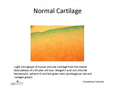
Normal Cartilage
-Light micrograph of human articular cartilage
from the medial tibial plateau of a 45-year old
man. Weigerts acid iron chloride hematoxylin,
safranin-O and fast green stain (proteoglycan red
and collagen green)
Provided by Y Henrotin
2
Normal cartilage
B
A
C
- Normal cartilage with chondrocytes isolated in an
extended - extracellular matrix (A). The calcified cartilage
layer overlying subchondral bone - (B). Normal subchondral bone (C).
Provided by Y Henrotin
3
Chondrocyte clusters in OA cartilage
- Chondrocyte clusters (arrows) are visible in the
fibrillated - superficial layer of OA cartilage
Provided by Y Henrotin
4
Subchondral bone sclerosis
- Subchondral bone sclerosis (arrow) is
characterized by trabecular - thickening, low mineralized osteoid substance
accumulation and - osteoblasts phenotype alteration
Provided by Y Henrotin
5
Multiple Tidemark
- Multiple tidemark are visible in OA calcified
cartilage (multiple arrows)
Provided by Y Henrotin
6
Osteochondral plate microcracks
A
- Subchondral bone sclerosis (A). Multiple
microcracks - are visible at the bone/cartilage junctions
(arrows)
Provided by Y Henrotin
Recommended
CrystalGraphics Presentations


![[PDF] OSTEOARTHRITIS FOR NEWLY DIAGNOSED: The complete clinical guide on the causes, symptoms and treatment of Osteoarthritis Full PowerPoint PPT Presentation](https://s3.amazonaws.com/images.powershow.com/10084247.th0.jpg?_=20240723114)


