addisa - PowerPoint PPT Presentation
Title: addisa
1
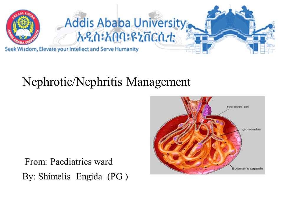
- Nephrotic/Nephritis Management
- From Paediatrics ward
- By Shimelis Engida (PG )
2
Presentation outlines
- Introduction
- Epidemiology
- Risk factors Aetiology
- Pathophysiology
- Clinical presentation
- Complications
- Treatment
- Evaluation and monitoring
3
Introduction
- The nephrotic syndrome is caused by renal
diseases that increase the permeability across
the glomerular filtration barrier. - Nephrotic range proteinuria -Urinary protein
excretion greater than 50 mg/kg per day - Hypoalbuminemia - Serum albumin concentration
less than 3 g/dL (30 g/L) - Oedema
- Hyperlipidaemia
4
(No Transcript)
5
Cont..
- The nephritic syndrome is a clinical syndrome
that presents as- - Haematuria
- elevated blood pressure
- decreased urine output, and oedema.
- The major underlying pathology is inflammation of
the glomerulus that results in nephritic syndrome
6
(No Transcript)
7
Epidemiology
- As per the final report by the National Center of
Health Statistics, nephritis syndrome, along with
nephrotic syndrome, is the 9th leading cause of
death in the USA in the year 2017. - The reported number of combined deaths due to the
nephritic syndrome, nephrotic syndrome, and renal
diseases was 50,633 out of a total of 2,813,503
deaths in the year 2017
8
Risk factors
- Risk factors of development of minimal change
disease include - Children within the Age gt1 year but lt8 years
- Hodgkin lymphoma
- Leukemia
- Recent viral illness
- Toxins such as mercury, gold, bee stings, fire
coral exposure. - Medication
9
Co
- focal segmental glomerulosclerosis (FSGS)
- Male gender
- Black race
- Family history
- Heroin abuse
- Drugs known to be associated with FSGS
- Chronic viral infection
- Single kidney status
- Obesity
- The following are considered risk factors for the
development of nephrotic syndrome
10
Etiology
- 90 - are primary glomerular abnormality
- Other are involvement of other disease (10)
- We can classify based on etiology
- Primary
- Secondary
- congenital and infantile nephrotic syndrome
11
Conti.....
- Based on pathological
- Minimal change disease (MCD)
- Focal segmental glomerulosclerosis (FSGS)
- Membranous glomeruli nephropathy
- Membranoproliferative glomerulonephritis (MPGN)
- Mesangial proliferation
- Focal and global glomerulosclerosis
12
Tendencies of Glomerular Diseases to Manifest
Nephrotic and Nephritic Features
13
CLASSIFICATION
- Primary nephrotic syndrome, which refers to
nephrotic syndrome in the absence of an
identifiable systemic disease. - Secondary nephrotic syndrome, which refers to
nephrotic syndrome in the presence of an
identifiable systemic disease. - Congenital and infantile nephrotic syndrome,
which occur in children less than one year of age
and can be either secondary (mostly due to
infection) or primary.
14
Pathophysiology
- Damaged glomerular capillary membrane
- Increase permeability of glomerular capillary
wall which leads to massive proteinuria, and
hypoalbuminemia - Decrease oncotic pressure
- Generalized edema
- Activation of RAAS
- Sodium retention edema
15
Con..
16
Clinical presentations
- Nephrotic
- Edema
- Weight gain
- Fatigue
- BP normal/raised
- Leukonychia
- Breathlessness
- Pleural effusion, fluid overload, AKI
- DVT/PE/MI
- Eruptive xanthomata/ xanthalosmata
- Nephritic
- Haematuria (E.g. colacoloured)
- Proteinuria
- Hypertension and edema as renal function declines
- Oliguria
- Flank pain
- General systemic symptoms
- Post-infectious 2-3 weeks after
strep-throat/URTI
17
Complications
- Malnutrition
- Nephrotic edema
- Infection of NS
- Thromboembolic complication
- Lipid abnormality
- Acute renal failure
- Chronic kidney disease
18
TREATMENT
19
Non-pharmacologic Therapy
- Restriction of sodium intake to 50 to 100 mEq/day
- To control edema, hypertension and proteinuria.
- Restriction of protein intake of 0.8 to 1 g/day
- To reduce proteinuria and progression of renal
disease - a low-fat diet of lt 200 mg cholesterol/day. Total
fat should account for lt 30 of daily total
calories. - plasmapheresis or plasma exchange, may be used to
remove the inflammatory mediators
20
Pharmacological Treatment
- Initial treatment of NS in children
- We recommend that oral glucocorticoids be given
for 8 weeks (4 weeks of daily glucocorticoids
followed by 4 weeks of alternate-day
glucocorticoids) or 12 weeks (6 weeks of daily
glucocorticoids followed by 6 weeks of
alternate-day glucocorticoids) (1B)
21
- Immunosuppressive Agents
- are commonly used to alter the immune processes
that are responsible for several of the
glomerulonephritides. - Corticosteroids reduce the production and/or
release of many substances that mediate the
inflammatory process - such as prostaglandins, leukotrienes,platelet-act
ivating factors, tumor necrosis factors, and
interleukin-1 (IL-1). - Cytotoxic agents, such as cyclophosphamide,
chlorambucil, or azathioprine, are commonly used
to treat glomerular diseases.
22
- The standard dosing regimen for the initial
treatment of nephrotic syndrome is daily oral
prednisone/prednisolone 60 mg/m2/d or 2 mg/kg/d
(maximum 60 mg/d) for 4 weeks followed by - alternate day prednisone/prednisolone, 40 mg/m2
or 1.5 mg/kg (maximum of 50 mg) for other 4
weeks, - or prednisone/prednisolone 60 mg/m2/d (maximum 60
mg/d) for 6 weeks followed by alternate day
prednisone/prednisolone, 40 mg/m2 or 1.5 mg/kg
(maximum of 50 mg), for other 6 weeks.
23
Prevention and treatment of relapses of NS in
children
- For children with frequently relapsing and
steroid-dependent nephrotic syndrome - If frequent relapse (2 or more relapses in the
initial 6 months or more than 3 relapses in any
12 months), - prednisolone 60mg/m2 (maximum 80mg) daily until
urinary protein turns negative or trace for 3
consecutive days followed by alternate day
therapy with 0.1-0.5mg/kg for 6 months and then
taper.
24
- For children with frequently relapsing nephrotic
syndrome who develop serio glucocorticoid-related
adverse effects and for all children with
steroid-dependent nephrotic syndrome - we recommend that glucocorticoid-sparing agents
be prescribed, rather than no treatment or
continuation with glucocorticoid treatment alone
(1B).
25
- If infrequent relapse (lt 2 relapses in 6 months
or lt 3 relapses in one year) - prednisolone 60mg/m2 (maximum 80mg) daily until
urinary protein turns negative or trace for 3
consecutive days followed by alternate day
therapy with 40mg/m2 (maximum 60mg) for 28 days
or 14 doses. - If the child relapses while on alternate day
prednisolone, - add levamisole 2.5mg/kg on alternate days for
6-12 months, then taper prednisolone and continue
levamisole for 2-3 years.
26
Steroid-resistant nephrotic syndrome in children
- We recommend using cyclosporine or tacrolimus as
initial second-line therapy for children with
steroid-resistant nephrotic syndrome (1C).
27
- Management of complication
28
Diuretics
- Management of nephrotic edema involves salt
restriction, bed rest, and use of support
stockings and diuretics. - Large doses (160 to 480 mg of furosemide)may be
needed for patients with moderate edema. - presence of large amounts of protein in the urine
promotes drug binding, and thereby reduces the
availability of the diuretic to the luminal
receptor sites. - reduced sodium delivery to the distal tubule
secondary to decreased glomerular perfusion may
also alter diuretic effectiveness. - thiazide diuretic or metolazone may be added to
enhance natriuresis.
29
Cont
- Alternatively, continuous IV infusion of a loop
diuretic, such as furosemide 160 to 480 mg/day,
may be employed. - For patients with morbid edema, albumin infusion
may be used to expand plasma volume and increase
diuretic delivery to the renal tubules - However, it may precipitate CHF and may
alsoreduce therapeutic response to steroids in
patients with minimal-change nephropathy. - For patients with significant edema, the goal of
treatment should be - a daily loss of 1 to 2 lb (0.45-0.9 kg) of fluid
until the patients desired weight has been
obtained.
30
Antihypertensive Agents
- Optimal control of hypertension for patients with
glomerular disease is important in reducing both
the progression of renal disease and the risk for
cardiovascular - According to JNC 8 guidelines, the target BP for
patients with CKD (GFRlt 60 mL/min/1.73 m2 is lt
140/90 mmHg - ACEIs ARBs delay the loss of renal function for
patients with diabetic and nondiabetic (primarily
glomerulonephritis)renal diseases. - Non-dihydropyridine calcium channel blockers (eg,
diltiazem and verapamil) reduce proteinuria and
preserve renal function and could be used as an
additional agent.
31
Antiproteinuria Agents
- ACEIs ARBs
- The antiproteinuric effect of ACEIs is associated
with a fall in filtration fraction, suggesting a
reduction in intraglomerular pressure. - ACEIs and ARBs may also have direct effects on
podocytes, resulting in reduction of proteinuria
and glomerular scarring. - ACEI inhibition may also reduce the effect of
angiotensin II on renal cell proliferation,
thereby reducing sclerosis. - ACEIs ARBs can reduce proteinuria through
differentmechanisms and combined use has been
shown to be more effective than monotherapy. - However, the risk of combination therapy has
become a concern recently.
32
Antiproteinuria Agents
- NSAIDs probably reduce proteinuria through PG E2
inhibition, resulting in a reduction of
intraglomerular pressure - Indomethacin and meclofenamate, the two most
evaluated NSAIDs have similar efficacy to ACEIs,
and combined - treatment with an ACEI results in
additionalproteinuria reduction. - Adherence to a low-sodium diet or concurrent use
of a diuretic is needed to maximize the
antiproteinuric effect. - Because of their potential for nephrotoxicity,
especiallyfor patients with preexisting CKD
33
Statins
- (HMG-CoA) reductase inhibitors, also known as
statins - such as lovastatin, pravastatin, simvastatin,
fluvastatin atorvastatin and rosuvastatin, - statins should be used to treat the dyslipidemia
- Aside from the lipid-lowering effects, statins
can reduce cardiovascular risk independent of
serum lipid concentrations. - Meta-analysis of published studies showed that
statins appear to reduce renal function decline
andslow the progression of proteinuria
moderately. - The beneficial effect may be dose-related
andduration-dependent
34
Anticoagulants
- Renal vein thrombosis andpulmonary emboli are
serious and common complications of nephrotic
syndrome, - patients who have documented thromboembolic
episodes should beanticoagulated with warfarin
until remission of nephrotic syndrome, - The use of prophylactic anticoagulation is
controversial. - A decision analysis study suggested that
prophylactic anticoagulation is beneficial for
patients with membranous nephropathy. - Prophylactic anticoagulation is recommended for
pts at high risk (ie, those with severe
nephrotic syndrome and a serum albumin
concentration less than 2-2.5.
35
Monitoring Parameters
- Renal function
- Serum creatinine concentration
- 24-h urine collection for creatinine clearance
determination - 24-h urine collection for urinary protein
excretion - Urine protein-to-creatinine ratio
- Clinical signs and symptoms
- Nephrotic syndrome
- Protein
- uriaSerum lipid concentrations
- Edema
36
- Nephritic presentations
- Hematuria
- Urinalysis
- Complete blood count
- Blood pressure
- General well-being appetite, energy levelKidney
biopsy to assess disease progression and response
to therapy - Assessment of drug therapy adverse reactions and
toxicities
37
Reference
- Heron M. Deaths Leading Causes for 2017. Natl
Vital Stat Rep. 2019 Jun68(6)1-77. PubMed


