Pulmonary cancer - PowerPoint PPT Presentation
Title:
Pulmonary cancer
Description:
The contains information about pulmonary cancer – PowerPoint PPT presentation
Number of Views:136
Title: Pulmonary cancer
1
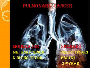
- PULMONARY CANCER
- MODERATOR SPEAKER
- Mr. ankit singh Preeti tiwari
- Nursing tutor Bsc (N)
-
3rd year
2
- LAYOUT
- Review of anatomy and physiology
- Introduction
- Historical aspect
- Incidence
- Definition
- Types of lung cancer SCLC
- NSCLC
- Stages of cancer
- Etiology
- Patho - physiology
- Signs and symptoms
- Diagnosis ,staging and grading
- Management Medical management
- Surgical management
- palliative care
- Nursing management
- Conclusion
- References
3
Review of anatomy and physiology of lungs
4
Anatomy
- The lungs are paired, elastic structures enclosed
in the thoracic cage , which is an right chamber
with distensible walls . - Weight of right lung 375 - 500gm
- Weight of left lung 325 -450 gm
- Each lung is divided into lobes .the right lung
has upper ,middle and lower lobes ,whereas the
left lung consists of upper and lower lobes. - Each lobe is further sub divided into 2-5
segments separated by fissures, which are
extension of pleura.
5
(No Transcript)
6
Pleura
- The lungs and thoracic cavity is lined with a
serous membrane called the pleura. - Pleura
- Visceral Parietal
- pleura pleura
- Pleural fluid(filled between
- these membranes)
- Mediastium
- Its in middle of thorax contain the 2 lungs.
- Alveoli
- Air filled sac like structure
- Approximately 300 million alveoli
7
Types of epithelial cells
8
Physiology of lungs
- Inhalation and exhalation are pulmonary
ventilation-thats breathing - External respiration exchanges gases between the
lungs and the bloodstream - Internal respiration exchanges gases between the
bloodstream and body tissues - Air vibrating the vocal cords creates sound
9
Introduction
- Lung cancer cells have accumulated a number of
molecular genetic and epigenetic lesions, which
appear necessary to transform normal bronchial
epithelium to an overt lung cancer . - Of the three major classes of human cancer
genes, the proto oncogenes and tumor suppressor
genes (TSGs) are involved in lung carcinogenesis
. - Tumors of the lung may be benign or malignant .
A malignant chest tumor can be primary ,arising
within the lungs ,chest wall or mediastinum or it
can be a metastasis from primary tumor site
elsewhere in the body . - Many of the proto oncogene and TSG changes are
present in both major lung cancer subtypes small
cell lung cancer (SCLC) and nonsmall cell lung
cancer (NSCLC) .
10
Historical aspects
- Lung cancer is the second highest cancer
incidence in both sexes after prostatemales
and breast females cancers . - It wasnt even recognized as a distinct disease
until 1761 . - In Germany in 1929 ,physician Fritz Lickint
recognized the between smoking and lung cancer
,which to an aggressive antismoking campaign . - First successful pneumonectomy was performed in
1933. - Palliative radiotherapy used since 1940s .
- Radical radiotherapy initially used in 1950s
11
Incidence
- Lung cancer mainly occurs in older people. About
2 out of 3 people diagnosed wit lung cancer are
65or older . - About 14 of all new cases of cancers are lung
cancer . - About 2,24,390 new cases of lung cancers1,17,920
in men and 1,06,470 in women . - Second highest cancer incidence in both sexes.
- Lung cancer has poor prognosis .
12
Definition
- Lung carcinoma ,is a malignant lung tumor
characterized by uncontrolled cell growth in
tissues of the lung . - If left untreated ,this growth can spread
beyond the lung by the process of metastasis into
nearby tissues or other parts of the body.
13
Types of lung cancer
14
Small cell lung carcinoma SCLC
- In this classification, SCLC was divided into
three subtypes that consist of oat cell,
intermediate cell type, and combined oat cell
(SCLC combined with squamous or adenocarcinoma). - Accounts for 15 of cases
- Generally starts in of the larger breathing
tubes or arises in the central airways and
initially infiltrates the submucosa, gradually
obstructing the lumen by extrinsic or
endobronchial spread. - Spreads more quickly and aggressively
- Found mostly in heavy smokers
15
(No Transcript)
16
Non small cell lung cancer NSCLC
- Most common type
- About 80-85 are NSCLC
- Grows more slowly
17
Classification of NSCLC a) large cell
carcinoma
- 10-20 cases are of lung cancer
- It can occur in any part of the lung
- Tends to grow and spread faster
- Cavitation common
- Although the WHO classification subdivides this
group into giant cell and clear cell varieties. - Histological view
18
b) Adenocarcinoma
- Increasing in frequency .
- 40-50 of all lung cancers
- Clearly defined peripheral lesions
- Glandular appearance under a microscope
- Easily seen on a CXR
- Can occur in non smokes
- Slow metastatic in nature pts present with or
develop brain ,adrenal or bone metastasis - Histological view
19
c) Squamous cell or epidermoid carcinoma
- Moderate to poor differentiation
- 30 -40 of lung cancer
- Arise from bronchial epithelium (occur centrally
in the large bronchi) - Uncommon metastasis,slow growth
- Associated with smoking
- slow growth
- Not easily visualized on x-ray
- Histological view
20
Stages of cancer
21
Etiology
- Tobacco smoke ( Of the three major classes of
carcinogens in tobacco smoke (polycyclic aromatic
hydrocarbons, such as benzoapyrene
nitrosamines and aromatic amines), - Second hand smoke
- Genetic predisposition
- Chromosomal
abnormality( In SCLCs, losses from chromosomes
3p, 5q, 13q, and 17p predominates In NSCLCs,
deletions of 3p, 9p, and 17p, together with 7,
i(5)(p10), and i(8)(q10) are often seen.) - Protooncogenes and growth
stimulation (Protooncogene products include
several growth factor receptors, such as
epidermal growth factor receptor (EGFR), ERBB2,
KIT, and MET.) - Tumor suppressor genes
and growth suppression (p53 or TP53 , p16INK4A,
Cyclin D1 and Cyclin-Dependent Kinase-4 ,
Retinoblastoma Protein )
22
- Occupational exposure
asbestos, 28,29,30,31,32,33,34,35,36,37,38 and
39 radon 40,41,42,43 and 44bis(chloromethyl)ethe
r, polycyclic aromatic hydrocarbons, chromium,
nickel, and inorganic arsenic compounds.
Silicosis ,coal workers pneumoconiosis and
environmental exposure . - Certain dietary supplements
- ß-carotene supplements ,are at
high risk - Over 50 years of age
23
Pathophysiology
24
(No Transcript)
25
(No Transcript)
26
Sign and symptoms
- There are two types of sign and symptoms of lung
cancer - Localized (involves lung)
- Generalized (involves other areas throughout the
body)
27
Localized Sign and symptoms
- Persistent cough and fatigue
- Breathing difficulty ,stridor
- Blood in phlegm
- Frank hemoptysis
- Hoarseness ,hiccups
- Weight loss
- Chest pain and tightness
- Pleural effusion
- Rust coloured purulent sputum
28
Generalized sign and symptoms
- Bone pain
- Headaches , mental status changes or neurological
findings - Abdominal pain
- Elevated LFT ,enlarged liver
- GI disturbances (anorexia ,dysphagia , cachexia
), jaundice - Weight loss
- Hand and neck edema
29
Staging and grading ( TNM Classification)
30
TNM Classification system
T N M Extent of primary tumor Absence or presence of extent of regional lymph node metastasis Absence or presence of distant metastasis
Primary tumor(T) Tx To Tis T1,T2,T3,T4 Cant be expressed No evidence of primary tumor Carcinoma in situ Increasing size of primary tumor
Regional lymph nodes(N) Nx No N1,N2,N3 Cant be expressed No metastasis of lymph node Increasing involvement of regional of regional lymph nodes
Distant metastasis(M) Mx Mo M1 Cant be assessed No distant metastasis Distant metastasis
31
Diagnostic evaluation
- History collection
- Physical examination
- CTX
- Sputum cytology
- Endoscopic ultrasound (EUS)
32
- Bronchoscopy
- It can identify early
- mucosal changes suggestive
- of lung cancer.
- Thoracoscopy
- Video-assisted thoracoscopy has been used
in the diagnosis and staging of lung cancer.
Peripheral nodules can be identified and excised
using video-assisted
33
- Needle biopsy
- Fine needle aspiration biopsy(FNA)
- Core biopsy
- CT guided biopsy
34
Medical management
- The use of radiation, chemotherapy,
immunotherapy, percutaneous albation and
palliative care either are given alone or in
combination. - 1. Radiation therapy
- Also called as radiotherapy, penetrating
waves or particles such as X-rays ,? rays. - Purpose kill or damage cancer cells
- Types of radiation
- a)External beam radiation
- b)Internal beam radiation
- c)Sealed source radiation
- d)Unsealed source of radiation
35
2. Percutaneous albation
- Percutaneous image guided ablation is a minimally
invasive treatment that can be offered to
patients with early stage NSCLC or palliative
treatment for patients with meatstatic disease
includes radiofrequency ablation, cryoablation
and microwave ablation. - 3. Chemotherapy
- Treatment of cancer with anti-cancer drugs.
- Types
- a) Adjuvant chemotherapy
- b)Neoadjuvant chemotherapy
36
Drugs
37
Surgical management
- Lobectomy
- The entire lobe containing the
- tumor is removed.
- Pneumonectomy
- Removal of entire lung.
38
- Wedge resection
- Removal of small, wedge-shaped
- piece of lung tissue to remove a small
- tumor or to diagnose.
- Segmental resection
- Also known as segmentectomy ,removal
- of a part of the lungs larger than a wedge
- section, but smaller than a complete lobe.
- Both surgeries may also be referred to as a
- sub-lobar resection.
39
- Thoracotomy
- Its a surgical incision into
- the thorax.
- Thoracoplasty
- Its a repair of the thoracic cavity.
40
- Pulmonary resection
- Complete resection of tumor remains
- the best chance of cure.
- Decortication
- Removal of the surface layer,
- membrane or fibrous cover of an organ.
- Bronchoscopic laser therapy
- Remove the obstructing lesions.
41
Palliative therapy
- Palliative care, concurrent with standard
0ncologic care for lung cancer, should be
considered early in the course of illness for any
patient with metastatic cancer. - The place of palliative care within course of
illness - Diagnosis of serious illness
Death
Life-prolonging therapy Palliative care
Medicare hospice benefit
42
Complications
- Respiratory failure
- Diminished cardiopulmonary function
- Pulmonary fibrosis
- Pericarditis
- Myelitis
- Pneumomitis
43
Nursing diagnosis
- Ineffective airway clearance related to increased
tracheo-bronchial secretions and presence of
tumor as evidenced by persistent cough, dyspnea. - Altered breathing pattern related decreased lung
capacity as evidenced by increased respiratory
rate, unexplained dyspnea. - Acute pain related to metastasis of tumor tissue
as evidenced by facial expression and from pain
score scale. - Imbalanced nutritional status less than body
requirement related to anorexia as evidenced by
decreased by decreased bodily weight. - Anxiety related to lack of knowledge about
pulmonary cancer as evidenced by verbal
communication with the client.
44
Nursing management
- Airway control
- Assess the patency of air.
- Assess the respiratory status and provide high
fowlers position. - Smoking cessation
- Provide awareness information for smoking
cessation classes. - Management symptoms
- The nurse educates the patient and family about
the potential side-effects of specific
treatments. - Reducing fatigue
- Fatigue is devasting symptom that affects quality
of life in patient with cancer.
45
Conclusion
46
References
47
Thank you .


