
Wisdom tooth - PowerPoint PPT Presentation
1 / 32
Title: Wisdom tooth
1
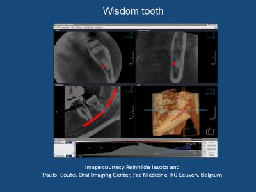
Wisdom tooth
Image courtesy Reinhilde Jacobs andPaulo Couto,
Oral Imaging Center, Fac Medicine, KU Leuven,
Belgium
2
Wisdom tooth
Image courtesy Reinhilde Jacobs andPaulo Couto,
Oral Imaging Center, Fac Medicine, KU Leuven,
Belgium
3
Wisdom tooth
Image courtesy Reinhilde Jacobs andPaulo Couto,
Oral Imaging Center, Fac Medicine, KU Leuven,
Belgium
4
Impacted canine
Image courtesy Reinhilde Jacobs andPaulo Couto,
Oral Imaging Center, Fac Medicine, KU Leuven,
Belgium
5
Impacted canine
Image courtesy Reinhilde Jacobs andPaulo Couto,
Oral Imaging Center, Fac Medicine, KU Leuven,
Belgium
6
Impacted canine and root resorption
Image courtesy Reinhilde Jacobs andPaulo Couto,
Oral Imaging Center, Fac Medicine, KU Leuven,
Belgium
7
Mesiodens
Image courtesy Reinhilde Jacobs andPaulo Couto,
Oral Imaging Center, Fac Medicine, KU Leuven,
Belgium
8
Mesiodens
Image courtesy Reinhilde Jacobs andPaulo Couto,
Oral Imaging Center, Fac Medicine, KU Leuven,
Belgium
9
Dentigerous Cyst
Image courtesy Reinhilde Jacobs andPaulo Couto,
Oral Imaging Center, Fac Medicine, KU Leuven,
Belgium
10
Dentigerous cyst
Image courtesy Reinhilde Jacobs andPaulo Couto,
Oral Imaging Center, Fac Medicine, KU Leuven,
Belgium
11
Infected cystic lesion
Image courtesy Reinhilde Jacobs andPaulo Couto,
Oral Imaging Center, Fac Medicine, KU Leuven,
Belgium
12
Periapical lesion
Image courtesy Reinhilde Jacobs andPaulo Couto,
Oral Imaging Center, Fac Medicine, KU Leuven,
Belgium
13
Periapical lesion
Image courtesy Reinhilde Jacobs andPaulo Couto,
Oral Imaging Center, Fac Medicine, KU Leuven,
Belgium
14
Periapical lesion
Image courtesy Reinhilde Jacobs andPaulo Couto,
Oral Imaging Center, Fac Medicine, KU Leuven,
Belgium
15
Root fracture
Image courtesy Reinhilde Jacobs andPaulo Couto,
Oral Imaging Center, Fac Medicine, KU Leuven,
Belgium
16
Root fracture
Image courtesy Reinhilde Jacobs andPaulo Couto,
Oral Imaging Center, Fac Medicine, KU Leuven,
Belgium
17
Root fracture
Image courtesy Reinhilde Jacobs andPaulo Couto,
Oral Imaging Center, Fac Medicine, KU Leuven,
Belgium
18
Root fracture with periradicular lesion
Image courtesy Reinhilde Jacobs andPaulo Couto,
Oral Imaging Center, Fac Medicine, KU Leuven,
Belgium
19
External cervical resorption
Image courtesy Reinhilde Jacobs andPaulo Couto,
Oral Imaging Center, Fac Medicine, KU Leuven,
Belgium
20
Cleft palate
Image courtesy Reinhilde Jacobs andPaulo Couto,
Oral Imaging Center, Fac Medicine, KU Leuven,
Belgium
21
Osteonecrosis in maxilla Osteoesclerosis in
mandible
Image courtesy Reinhilde Jacobs andPaulo Couto,
Oral Imaging Center, Fac Medicine, KU Leuven,
Belgium
22
Salivary gland stone
Image courtesy Reinhilde Jacobs andPaulo Couto,
Oral Imaging Center, Fac Medicine, KU Leuven,
Belgium
23
Aggressive Periodontal Disease
Image courtesy Reinhilde Jacobs andPaulo Couto,
Oral Imaging Center, Fac Medicine, KU Leuven,
Belgium
24
Condensing Osteitis
Image courtesy Reinhilde Jacobs andPaulo Couto,
Oral Imaging Center, Fac Medicine, KU Leuven,
Belgium
25
Condensing Osteitis
Image courtesy Reinhilde Jacobs andPaulo Couto,
Oral Imaging Center, Fac Medicine, KU Leuven,
Belgium
26
Enostoses
Image courtesy Reinhilde Jacobs andPaulo Couto,
Oral Imaging Center, Fac Medicine, KU Leuven,
Belgium
27
Bone defect
Image courtesy Reinhilde Jacobs andPaulo Couto,
Oral Imaging Center, Fac Medicine, KU Leuven,
Belgium
28
Atelectasis of the maxillary sinus
Image courtesy Reinhilde Jacobs andPaulo Couto,
Oral Imaging Center, Fac Medicine, KU Leuven,
Belgium
29
Sinusitis
Image courtesy Reinhilde Jacobs andPaulo Couto,
Oral Imaging Center, Fac Medicine, KU Leuven,
Belgium
30
Sinusitis
Image courtesy Reinhilde Jacobs andPaulo Couto,
Oral Imaging Center, Fac Medicine, KU Leuven,
Belgium
31
Buccosinusal fistula
Image courtesy Reinhilde Jacobs andPaulo Couto,
Oral Imaging Center, Fac Medicine, KU Leuven,
Belgium
32
Nasopalatine cyst
Image courtesy Reinhilde Jacobs andPaulo Couto,
Oral Imaging Center, Fac Medicine, KU Leuven,
Belgium
Recommended
CrystalGraphics Presentations


