Ankle - PowerPoint PPT Presentation
1 / 30
Title:
Ankle
Description:
Calcaneus, Talus, Tibia (Medial Malleolus), Fibula (Lateral Malleolus) Soft Tissue ... Bump (Tap or Percussion) test (stress fracture) ROM AROM, PROM, RROM ... – PowerPoint PPT presentation
Number of Views:114
Avg rating:3.0/5.0
Title: Ankle
1
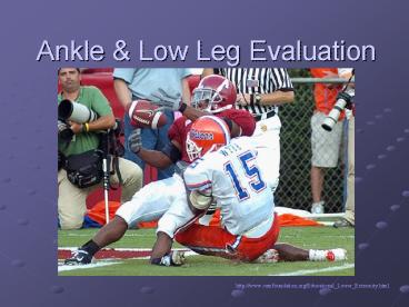
Ankle Low Leg Evaluation
http//www.csmfoundation.org/Educational_Lower_Ext
remity.html
2
Common Injuries
- Ankle sprains lateral, medial, syndesmosis
(high) - Fractures
- Tendinitis
- Subluxating Peroneal Tendons
- Compartment Syndromes (Anterior)
- Achilles Tendon Rupture
- Deep Vein Thrombosis (Thrombophlebitis)
3
Ankle Anatomy
- Bony
- Calcaneus, Talus, Tibia (Medial Malleolus),
Fibula (Lateral Malleolus) - Soft Tissue
- Muscles
- Ligaments
- Interosseous Membrane
- Retinaculum
- Peroneal, Extensor (superior, inferior)
- Circulation
- Posterior Tibial Artery, Anterior Tibial Artery,
Peroneal Artery, Veins
4
Joints Ligaments
- Medial Ligaments
- Deltoid ligament 2 layers (deep superficial)
- Superficial 3 parts, Deep 1 part
5
Joints Lateral Ligaments
- Anterior Talofibular Lig. (ATF)
- Anterior Tibiofibular Lig.
- Posterior Talofibular Lig.
- Posterior Tibiofibular Lig.
- Calcaneofibular Lig.
6
Joints
- Talocrural Joint dorsi plantarflexion
- Subtalar Joint - inversion eversion
- Classified as gliding or arthrodial
- Intertarsal tarsometatarsal joints
- Arthrodial
- Minimal movement
7
Muscles of the Ankle Low Leg
- Anterior Compartment
- Tibialis Anterior
- EHL
- EDL
- Peroneus Tertius
- Lateral Compartment
- Peroneus Longus
- Peroneus Brevis
- Superficial Posterior Compartment
- Gastrocnemius
- Soleus
- Plantaris
- Deep Posterior Compartment
- Tibialis Posterior
- FHL
- FDL
8
Anterior Leg Muscles
9
Lateral Medial Leg Muscles
10
Posterior Leg Muscles - Deep
11
Posterior Leg Muscles - Superficial
12
Thanks Katie!
- MAP(L) OF SCIATIC(L)
- Medial Obturator Nerve
- Anterior Femoral Nerve
- Posterior Sciatic Nerve
- (L)ateral (L)ateral Cutaneous Nerve
13
Neurological Anatomy - Posterior
14
Nerves
- Sciatic nerve
- Tibial division
- gastrocnemius (medial head)
- soleus
- tibialis posterior
- flexor digitorum longus
- flexor hallucis longus
- Medial plantar nerve
- Lateral plantar nerve
15
Nerves
- Sciatic nerve
- Common peroneal (fibular) division
- Superficial peroneal nerve
- peroneus longus
- peroneus brevis
- Deep peroneal nerve
- tibialis anterior
- extensor digitorum longus
- extensor hallucis longus
- peroneus tertius
- extensor digitorum brevis
16
Neurological Anatomy - Anterior
17
Nerves
- Femoral nerve
- Iliopsoas
- Rectus femoris
- Vastus medialis
- Vastus intermedius
- Vastus lateralis
- Pectineus
- Sartorius
- Saphenous nerve
- Medial leg foot
18
Nerves
- Obturator Nerve
- Adductor brevis
- Adductor longus
- Adductor magnus
- Gracilis
- Obturator externus
- Sensation to medial thigh
19
Neurovascular Anatomy of Leg
20
Dermatomes (L4 green, L5 purple, S1 flesh)
21
Movements
- Eversion
- Turning ankle foot outward abduction, away
from midline weight is on medial edge of foot - 5-15º of eversion
- Inversion
- Turning ankle foot inward adduction, toward
midline weight is on lateral edge of foot - 20-30º of inversion
- Dorsiflexion (flexion)
- Movement of top of ankle foot toward anterior
tibia - 15-20º of dorsiflexion
- Plantar flexion (extension)
- Movement of ankle foot away from tibia
- 50º of plantar flexion
22
Evaluation of the Ankle Low Leg
- History
- What happened? (MOI)
- Where is the pain?
- What causes the pain?
- When did it happen? (onset)
- Has it happened before?
- What does it feel like?
- Pain scale (1-10)
- What type of surface?
- How old are the shoes?
- Type of pain
- Unusual noises/sensations
23
What is the History? What do you See?
24
Observation
- Appearance
- How is their ankle hanging?
- Bilateral comparison
- Color
- Deformity
- Edema/Swelling
- Is there a lot of swelling immediately?
- Gait
- Infection
- Weight bearing vs. non-weight bearing
25
What Do You See?
26
Evaluation of the Ankle Low Leg
- Palpation
- Start away from the point of pain
- Medial structures
- Lateral structures
- Dorsal structures
- Plantar structures
- Crepitus
- Heat
- Swelling
- Rigidity
- Deformities
- Stress/ Special Tests
- ROM tests (AROM, PROM, RROM-strength)
- Special Tests
- Fracture tests
- Ligament/Capsular tests
- Neurologic tests
- Other
Which stress/special test do you perform first?
How do you make the correct decision?
27
Stress/ Special Tests
When do you perform all of these tests?
- Lower Leg Fractures
- Compression (Squeeze) test (fracture)
- Bump (Tap or Percussion) test (stress fracture)
- ROM AROM, PROM, RROM
- Talar Tilt (Inversion Eversion Stress) tests
(calcaneofibular ligament or deltoid ligament
instability) - Anterior Drawer test (ATF ligament instability)
- Kleigers Test (syndesmosis instability)
- Homans Sign (deep vein thrombophlebitis
neurovascular pathology) - Thompson test (achilles tendon pathology)
28
Whats your Diagnosis?
- What is the MOI?
- Where is the pain elicited?
- Which tests elicit positive findings?
- What are your observations?
- How is the ROM strength?
- Are there any neurovascular symptoms?
- What do you think the injury is?
29
What Do You Do Next?
- Is referral needed?
- RICES
- Are any home instructions
- needed?
30
Conclusion
- Questions?????
- Case Study
- Trivia
- What is the os calcis?
- What is an os trigonum injury?


