Advanced Airway Techniques - PowerPoint PPT Presentation
1 / 42
Title:
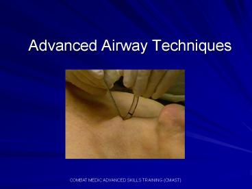
Advanced Airway Techniques
Description:
Casualty vomits past either distal or pharyngeal tube ... Insert the end of the ET tube into the trachea directed towards the lungs and ... – PowerPoint PPT presentation
Number of Views:1329
Avg rating:3.0/5.0
Title: Advanced Airway Techniques
1
Advanced Airway Techniques
COMBAT MEDIC ADVANCED SKILLS TRAINING (CMAST)
2
Introduction
- One of the most critical skills for the soldier
medic. - Without proper airway management and ventilation
techniques, casualties may die. - Must be able to choose and effectively utilize
the proper equipment for ventilation in a
tactical environment.
3
Review the Physiology
- Inhalation (an active process)
- Initiated by contracting of respiratory system
muscles - Diaphragm contracts and drops downward
- Intercostal muscles contract, chest expands
- Intrathoracic pressure falls, pulling air into
lungs - Exhalation (a passive process)
- Respiratory muscles relax diaphragm moves upward
- Chest wall recoils
- Intrathoracic pressure rises
- Air is pushed out
4
Gas Exchange
- Alveoli supply O² to, and remove CO² from the
lungs. - Exchange is made by diffusion across the cell
wall of the alveoli and capillaries.
Inhalation
Exhalation
5
Sources of Airway Obstruction
- Tongue
- Most common cause of airway obstruction
- Foreign body airway obstruction (FBAO).
- Trauma/Combat
- Loose teeth, facial bone fractures, fractured
larynx - Laryngeal spasm
- Edema can severely obstruct airflow
- Aspiration.
6
Nasopharyngeal Airway
- Insert a nasopharyngeal airway (NPA) adjunct.
7
Nasal Airway Adjunct
- Do not use if roof of mouth is fractured or brain
matter is exposed. - Purpose
- To maintain an artificial airway for oxygen
therapy or airway management
8
Nasal Airway Adjunct
- Indications
- Conscious, semi-conscious or has an active gag
reflex - Injuries to mouth
- Seizure casualties
- Likely vomiting
9
Nasal Airway Adjunct
- Contraindications
- Injuries to roof of mouth
- Exposed brain matter
- Drainage of CSF from nose, mouth or ears
10
Nasal Airway Adjunct
- Complications
- Nasal trauma
- Bloody nose, minor tissue trauma (most common)
- May trigger gag reflex if NPA is too long
11
Nasopharyngeal Insertion
- Procedures
- Supine position on firm surface C-spine
stabilized - Select proper size NPA
- Diameter smaller than the casualtys nostril
approximately diameter of casualtys little
finger - Length - Measure from tip of nose to earlobe
12
Nasopharyngeal Insertion
- Procedures
- Lubricate the NPA with a water soluble lubricant
13
Nasopharyngeal Insertion
- Procedures
- Place head into a neutral position extend
nostril
14
Nasopharyngeal Insertion
- Procedures
- Insert tip of the NPA through the R nostril if
resistance is met,
do not force, try - the other nostril
- Place casualty
- In recovery
position
15
Combitube
- Esophageal-tracheal double
lumen airway. - Blind insertion.
- Successful in casualties with
- Trauma
- Upper airway bleeding and
vomiting - Effective in cardiopulmonary
resuscitation.
16
Combitube
- Double-lumen design allows for effective
ventilations regardless if in the trachea or
esophagus. - Comes in two sizes
- 37 Fr
- 41 Fr
17
Combitube
- Indications
- Adult casualties in respiratory distress
- Adult casualties in cardiac arrest
- Contraindications
- Intact gag reflex
- Casualties less than 5 feet in height
- Known esophageal disease
- Caustic substance ingestion
18
Combitube
- Side effects and complications
- Sore throat
- Dysphagia
- Upper airway hematoma
- Esophageal rupture (rare).
- Preventable by avoiding over-inflation of the
distal and proximal cuffs.
19
Combitube
- Intubation procedures
- Inspect the upper airway for visible obstructions
- Hyperventilate ( 20/min) for 30 seconds
- Casualty in neutral
head position - Test both cuffs
- 15 ml (white)
- 100 ml (blue)
20
Combitube
- Intubation procedures
- Insert in same direction as the natural curvature
of the pharynx - Grasp tongue and lower jaw between thumb and
index finger, lift upward (jaw-lift) - Insert gently but firmly until black rings are
positioned between casualtys teeth - Do not force if does not insert easily,
withdraw and retry - Hyperventilate between attempts
21
Combitube
- Intubation procedures
- Inflate 1 (blue) pilot balloon with 100 ml of
air (100 ml syringe) - Inflate 2 (white) pilot balloon with 15 ml of
air (20 ml syringe) - Ventilate through the
primary 1 blue tube if
auscultation of breath
sounds is positive
(gastric sounds is
negative), continue to ventilate
22
Combitube
- Intubation procedures
- If auscultation of breath sounds is negative and
gastric sounds is positive, immediately begin
ventilations through the shorter (white)
connecting tube (2) - Confirm tracheal ventilation
of breath sounds and
absence
of gastric insufflation
23
Combitube
- Intubation procedures
- If auscultation of breath sounds and auscultation
of gastric insufflation is negative, the
Combitube? may have been advanced too far into
the pharynx - Deflate the 1 balloon/cuff, and move the
Combitube? approx. 2-3 cm. out of the casualtys
mouth - Re-inflate the 1 balloon and ventilate through
the longer (1) connecting tube if auscultation
of breath sounds is positive and auscultation of
gastric insufflation is negative continue to
ventilate. - If breath sounds are still absent extubate
24
Combitube
- Combitube removal.
- Should not be removed unless
- Tube placement cannot be determined
- Casualty no longer tolerates the tube
- Casualty vomits past either distal or pharyngeal
tube - Palpable pulse and casualty breathing on their
own - Physician or PA is present to emplace ETT
25
Combitube
- Combitube removal.
- Have suction available and ready
- Logroll casualty to side (unless spinal-injured)
- Deflate the pharyngeal cuff (1 pilot balloon)
- Deflate the distal cuff (2 pilot balloon)
- Gently remove Combitube? while suctioning
26
Emergency Cricothyrotomy
- Indications
- Inability to ventilate a casualty with NPA or
Combitube secondary to - Severe maxillofacial injury, airway obstruction
and structural deformities - Emergency airway catheters with a 6 mm diameter
allow for spontaneous breathing and adequate
oxygenation in adults
27
Emergency Cricothyrotomy
- When maxillofacial, cervical spine, head or soft
tissue injuries are present, several factors may
prevent ventilation - Gross distortion
- Airway obstruction
- Massive emesis
- Significant hemorrhage
28
Emergency Cricothyrotomy
- Complications
- Incorrect tube placement
- Blood aspiration
- Esophageal laceration
- Hematoma
- Tracheal wall perforation
- Vocal cord paralysis, hoarseness
29
Larynx
30
Cricothyroid Membrane
Thyroid Cartilage
Cricothyroid Membrane
Cricoid Cartilage
31
Emergency Cricothyrotomy
- Procedure
- Identify and palpate the cricothyroid membrane
- Make a 1 ½-inch vertical incision in the midline
using a 15 or 10 scalpel blade
32
Emergency Cricothyrotomy
- Procedure
- Stabilize the larynx with one hand using a
scalpel or hemostat, cut or poke through the
cricothyroid membrane - A rush of air may be felt through the opening
33
Emergency Cricothyrotomy
34
Emergency Cricothyrotomy
- Insert the end of the ET tube into the trachea
directed towards the lungs and inflate the cuff
with 5-10 ml of air - Advance the tube no more than 2-3 inches further
intubation could result in right main stem
broncus inubation only
35
Emergency Cricothyrotomy
- Check for air exchange and tube placement
- Listen and feel for air passing in and out of
tube - Look for bilateral rise and fall of the chest
- Ascultate the abdomen and both lung fields
36
Emergency Cricothyrotomy
- Indications of proper placement
- Unilateral breath sounds and rise and fall of the
chest (right main stem intubation) deflate cuff
and retract 1-2 inches and recheck airway - Air coming out of the casualtys mouth (tube
pointing away from lungs) remove tube and
reinsert with tube facing lungs
37
Emergency Cricothyrotomy
- If casualty is not breathing spontaneously direct
someone to perform rescue breathing - Connect tube to BVM and ventilate at 20 breaths
per minute - No BVM available, perform mouth-to-tube
resuscitation at 20 breaths per minute - Tube must be secured once rescue breathing has
started
38
Emergency Cricothyrotomy
- Apply dressing to protect the tube and incision
site - Cut two 4x4 gauze sponges halfway through and
place on opposite sides of tube tape securely - Or apply two 4x4 gauze dressing in a V shape
fold at the edges of the cannula and tape
securely
39
Emergency Cricothyrotomy
- Monitor casualtys respirations on a regular
basis. - Reassess air exchange and tube placement every
time the casualty is moved - Assist with respirations if rate falls below 10
or above 24 per minute
40
Emergency Cricothyrotomy
Click in box for video
41
Summary
- Airway compromise is a small percentage of combat
casualties. - Airway management must be readily available and
rapidly applied. - Airway compromise is the third leading cause of
preventable death on the battlefield.
42
Questions?


