
What do these organisms have in common - PowerPoint PPT Presentation
1 / 15
Title:
What do these organisms have in common
Description:
What do these organisms have in common? Jellyfish (Aequorea victoria) Renilla reniformis ... These proteins are naturally found in light-producing cells of ... – PowerPoint PPT presentation
Number of Views:33
Avg rating:3.0/5.0
Title: What do these organisms have in common
1
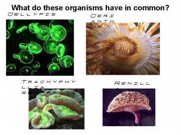
What do these organisms have in common?
Jellyfish (Aequorea victoria)
Cerianthus
Trachyphyllia geoffroyi
Renilla reniformis
2
They all contain fluorescent proteins
These proteins are naturally found in
light-producing cells of COLENTERATES
(JELLLYFISH, ANEMONES, CORALS, ETC.)
Red Fluorescent Protein (dimer)
Green Fluorescent Protein (monomer)
3
Why would a researcher use a fluorescent protein?
To visualize mouse metabolism and study
abnormalities.
4
Cancer cells can easily be observed and monitored
in living GFP mice.
- Mouse under blue light (left) Same mouse under
normal light (right)
Mouse blood vessels (green-GFP)) in tumor
(red-DsRed). Mouse with brain tumor expressing
DsRed.
5
Scientists can now visualize mitosis
What stage of mitosis is this? gt gt gt
6
Scientists can now clone FP to look at cell
structures
Which cell structures can you identify? Animal
or plant cells?
7
Looking at the cytoskeleton of a cell
Two different types of FPs are used to show
contrasting cell structures.
8
Looking at other types of cells
Plant or animal cell?
9
GFP in Sperm cells of Spiders
In most species of spiders, insects and birds,
multiple males mate with a single female. GFP
can determine which sperm cells actually
fertilize the egg.
10
- Other FP uses
- Screening drugs
- Evaluating viral vectors for human gene therapy
- Biological pest control
- Monitoring genetically altered microbes
11
How do scientists tag genes with FP?
- Suppose a researcher wants to study a protein of
interest, - First the gene for that protein is isolated.
- The gene is put in a cell via a vector (like a
plasmid). - The cell will go through transcription and
translation to make the protein. - But, how does the researcher detect the protein
created? - continue.
12
How do scientists tag genes with FP?
- The FP gene is inserted right after the gene for
the protein, before the stop codon. - The protein of interest AND the FP are copied and
translated together. - Now, the scientist can see the protein of
interest (its location) and measure the amount of
protein translated (how much it fluoresces a
particular color).
13
How plasmids are genetically engineered
DNA Plasmid Vector
Host DNA fragments (i.e. coral or jellyfish DNA
Ligate fragments into cut DNA vector
Cut plasmids open with DNA enzymes
Cut genomic DNA into fragments
End result Plasmid containing FP gene
14
We will be performing a transformation
- Transformation is the method of introducing the
plasmid vector to E. coli. - Two methods of transformation
- Heat shock and electroporation, we will use heat
shock.
Plasmid
Transformation
Allow bacteria to grow for 1-3 days on plate with
ampicillin.
Plasmid
Bacteria now express cloned fluorescent protein
(transcription of gene and translation of mRNA to
protein at ribosomes).
15
Why have an Amp(icllin) resistance gene in the
plasmid?
Transformation is NOT 100 effective Plating on
amp is one way to select for bacteria that have
been transformed. The Amp resistance gene codes
for an enzyme, Beta Lactamase. This enzyme
breaks up ampicillin.
Transformation step
Plasmid with FP and amp resistance
Tube of E.coli
Heat shock to insert plasmid
Plate transformed bacteria
Petri plate has nutrient agar and ampicillin
Recommended
CrystalGraphics Presentations


