Core II: Lecture 3: Cellular Differentiation - PowerPoint PPT Presentation
1 / 55
Title:
Core II: Lecture 3: Cellular Differentiation
Description:
of a given gene is not achieved by a single transcription. factor: unique combinations ... with each other when bound to the same promoter in cis; ... – PowerPoint PPT presentation
Number of Views:611
Avg rating:3.0/5.0
Title: Core II: Lecture 3: Cellular Differentiation
1
Core II Lecture 3 Cellular Differentiation
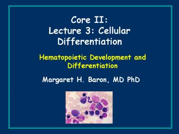
Hematopoietic Development and Differentiation
- Margaret H. Baron, MD PhD
2
(No Transcript)
3
Gene expression analyses suggest that stem cells
possess a wide open chromatin structure to
maintain their multipotentiality, which is
progressively restricted as they proceed down a
particular pathway of differentiation
4
More restricted progenitors
Multiplex single-cell RT-PCR lineage
promiscuity in CMPs and CLPs precedes lineage
commitment in hematopoiesis
Genes unrelated to the adopted lineage are
downregulated in bipotent and unipotent
descendents of CMPs and CLPs
Miyamoto et al. (2002) Dev. Cell 3137
5
Hierarchical distribution of hematopoietic stem
and progenitor cells
Common lymphoid progenitor
Follow-up study using microarrays
Multipotent progenitor
Common meloid progenitor
Stem and progenitor cells were prospectively
FACS-purified and their gene expression patterns
were then analyzed
Akashi et al. (2003) Blood 101383
6
Promiscuous expression of non-hematopoiesis-associ
ated genes in HSCs
i.e. HSCs co-express not only hematopoietic but
multiple non-hematopoietic genes
Akashi et al. (2003) Blood 101383
7
Summary of observations from these studies
MPPs co-express myeloid and lymphoid genes
CMPs co-express myelo-erythroid but not lymphoid
genes
CLPs co-express T-, B- and NK-lymphoid but not
myeloid genes
Stepwise decrease in transcriptional
accessibility for multilineage-associated genes
may represent progressive restriction of
developmental potentials during hematopoiesis
Akashi et al. (2003) Blood 101383
8
Clusters of genes categorized by expression
patterns in purified stem and progenitor cells
Representative genes predominantly expressed in
HSCs
MPPs
CLPs
CMPs
Akashi et al. (2003) Blood 101383
9
Hematopoietic and non-hematopoietic lineage
promiscuity
Common lymphoid progenitor
Common meloid progenitor
Multipotent progenitor
Stepwise decreases of lineage potentials and
lineage promiscuity during early hematopoiesis.
Lineage promiscuity is distributed in a
hierarchical and asymmetrical fashion.
Akashi et al. (2003) Blood 101383
10
(No Transcript)
11
Transcriptional control of hematopoiesis
general comments
Tissue-specific and developmentally correct
expression
of a given gene is not achieved by a single
transcription
factor
unique combinations
of cell-type specific and
widely-expressed nuclear factors account for
specificity
and diversity in gene expression profiles
in addition, most hematopoietic transcription
factors are
expressed in more than one cell type
12
Null mutations in certain hematopoietic
transcription factors may have different
effects on primitive vs. definitive
hematopoiesis or may affect both
13
Cis-acting regulatory elements
from Lemon and Tjian
14
Activators and the basal transcription machinery
from Lemon and Tjian
15
from Lemon and Tjian
Co-activators are required to mediate activator
responsiveness (TAFs discovered first, others
since then)
16
from Lemon and Tjian
Great diversity of co-regulators co-activators
and co-repressors mediate signals between
sequence-specific transcription factors and the
core transcriptional machinery
CBP/p300
FOG
17
Co-factors e.g. CREB-binding protein (CBP) and
p300
CBP and p300 function as "molecular integrators"
via protein-protein interactions
nuclear factors that interact with CBP and p300
can synergize
with each other when bound to the same promoter
in cis
however, inhibition between these factors might
occur if they are
bound to different promoters -- competition for
limiting amounts
of CBP or p300 in nucleus (e.g. STAT vs. AP-1)
18
Many protein interactions involving CBP and p300
are regulated by cellular signals
1. phosphorylation of CREB regulates interaction
with CBP
and p300
2. steroid hormones or retinoic acid (small
ligands) stimulate
CBP or p300 binding to nuclear hormone receptors
3. Acetylation by histone acetyl transferase
(HAT) or
de-acetylation by histone deacetylase (HDAC)
Epigenetic regulation of gene expression
19
Most transcriptional cofactors act
stoichiometrically
CBP, p300 have
enzymatic activities
that allow them to function
non-stoichiometrically -- action by catalysis
Intrinsic histone acetyltransferase (HAT)
activity may
facilitate access of transcription factors to DNA
20
Acetylation of transcription factors by CBP, p300
etc.
e.g. p53, EKLF, GATA-1, NF-E2
erythroid transcription factors controlling
globin gene expression
may cooperate in the formation of a high
molecular weight complex
in which GATA-1, NF-E2, and EKLF are linked
through CBP and p300
21
(No Transcript)
22
CBP and p300 may function in part by linking
DNA-bound nuclear factors to
components of the basal transcription machinery
(general transcription
factors, TATA-binding protein, TAFs, Pol II)
23
Acetylation may regulate
DNA binding by transcription factor
response to CBP or p300
interactions with other transcription/co-factors
changes in protein conformation, recruitment of
other transcription factors -- ?
24
GATA-1 is one of several DNA-binding
transcription factors that are acetylated by
p300/CBP
Modification of GATA-1 by HATs such as p300/CBP
occurs at two highly conserved lysine rich motifs
located near the zinc fingers
Acetylation may modulate biological activity of
GATA-1 but does not appear to affect DNA binding
specificity or affinity
Acetylation of GATA-1 may regulate changes in
conformation, function in recruitment of other
proteins (by providing a new docking site)
25
p300 contains a transcription factor-binding
surface that is required for hematopoiesis
p300 and CBP contain protein binding KIX domains
(among other domains) that have nonredundant
functions (gene targeting experiments)
Homozygous point mutations in KIX domain of p300
designed to interfere with binding to c-Myb and
CREB result in hematopoietic defects in multiple
lineages. Same mutations in CBP mice are
normal.
Synergistic genetic interaction between mutations
in c-Myb and mutations in KIX domain of p300
binding of c-Myb to this domain is critical.
Kasper et al. (2002) Nature 419738
26
Modification of chromatin not
restricted to acetylation
numerous regulated chromatin remodeling
complexes have been identified
SWI/SNF complex, an ATP-dependent
chromatin remodeling machine
27
Mutations in transcriptional regulators
in hematopoietic cells as one cause of disease
Point mutation in GATA1 that reduces DNA
binding and interaction with FOG1
family with severe dyserythropoietic anemia
ATRX and chromatin remodeling
a-thalassemia/X-linked mental retardation
translocations create transcription factors
with new activities leukemias
SCL/tal-1
AML1/cbfa2/runx1 (AML-ETO)
PLZF
28
(No Transcript)
29
Specificity of erythroid, myeloid, T cell etc.
promoters is mediated by combinatorial
interactions among transcription factors and
co-regulators in those cell types
Example M-CSF-receptor promoter in monocytes
CBFb
1
mRNA
CBFa/ AML1
PU.1
C
C/EBPa
In hematopoietic cells, C/EBPa is myeloid
specific (i.e. one subtype of white blood cells
but not all) AML1 is expressed in ALL white
blood cells, PU.1 is B- and myeloid-cell specific
major cell type in which all three
proteins are expressed is the myeloid cell
C/EBPa and AML1 interact and synergize to
activate promoter
On other promoters, C/EBP proteins can synergize
with PU.1
CBF??heterodimerizes with AML1, does not bind DNA
itself but strengthens/stabilizes DNA binding by
AML1
30
Lineage-specific transcription factors may
inhibit one another's activities
Depending on the cell type
cross-antagonism
GATA1 PU.1
composition and balance of transcription factors
within a cell is a critical determinant of cell
lineage/differentiation
PU.1 GATA1
FOG CEBPb
CEBPb FOG
31
Protein-protein interactions may alter DNA
binding in EMSA ( , , abolish, change
mobility of complex)
Protein-protein interactions may change activity
in trans-activation assays (e.g. luciferase
reporter)
Protein-protein interactions may change
biological activity in transfected cells (i.e.
establishment of a particular lineage/phenotype)
32
interact via ets domain
(a) GATA-1 mediated antagonism of PU.1
interact via ets domain ZF
interact via C-ZF and TAD
binds via C-ZF
(b) PU.1 mediated antagonism of GATA-1
Cross-antagonism of PU.1 and GATA-1
from Cantor and Orkin (2001) Curr. Opin. Genet.
Dev. 11513
33
(a) C/EBPb represses FOG gene expression at
transcriptional level
(b) FOG acts as transcriptional co-repressor of
some C/EBPb target genes through interactions
with GATA-1
Cross-antagonism between C/EBP-b and FOG
from Cantor and Orkin
34
Transcription factor complexes in the
erythroid/megakaryocyte compartment
More generally progression from stem/progenitor
cell to differentiated cell may involve
sequential protein complex formation (still
speculative)
from Sieweke and Graf (1998) Curr. Opin. Genet.
Dev. 8545
35
Cross-antagonistic transcription factor model of
lineage determination
from Cantor and Orkin
36
(No Transcript)
37
(No Transcript)
38
(e.g. ectopic expression of a transcription
factor)
39
(No Transcript)
40
Erythropoiesis as a model for
cellular differentiation
41
In general
42
primitive
LCR
4
3
2
1
5
g
g
b
e
d
G
A
e
m
b
r
y
o
f
e
t
u
s
a
d
u
l
t
definitive
stage-specific
regulators
Some of the transcriptional regulators required
for globin gene activation and silencing
("switching") and for regulation of expression of
other erythroid cell-specific genes are ALSO
required for formation and differentiation of red
blood cells.
43
(No Transcript)
44
(No Transcript)
45
(No Transcript)
46
Genotypes of viable progeny derived from
heterozygous parents
Genotype
Stage
Total
/ /- -/-
Postnatal 90
30 60 0
E13.5 12
4 8 0
E12.5 22
10 12 0
(0)
E11.5 156
43 91 22
(14)
E10.5 111
26 56 29
(26)
Viable embryos defined by presence of beating
heart.
KOs dies between E10.5 and E12.5 (loss of
Mendelian ratios)
47
FOG KO erythroid cells show marked, but partial
arrest in development at proerythroblast
stage Reminiscent of GATA1-deficient erythroid
progenitors
48
Peripheral blood cells
49
(No Transcript)
50
MGG May-Grunewald Giemsa stain
yolk sac, fetal liver cells
51
(No Transcript)
52
Erythroid block Megs also abnormal,
hyperproliferate
bipotential
progenitor
Erythroid block formation of Megs completely
blocked
53
NF-E2 a heterodimeric bZIP protein
p45 NF-E2 in erythroid cells, megakaryocytes (as
is GATA-1)
p18 small maf proteins, widely expressed
(family)
binds to several core sites within locus control
region (LCR) upstream from b-globin gene locus in
erythroid cells
gene targeting of p45 NF-E2 results in profound
defects in platelet formation but only mild
defects in erythroid lineage
are there other p45 NF-E2
family members in erythroid cells that could
compensate for its absence?
functional redundancy, protein families
54
In Summary The commitment of multipotent
stem/progenitor cells to particular developmental
pathways requires specific changes in their
complement of transcription factors to generate
the patterns of gene expression characteristic of
specialized cell types (erythroid, myeloid,
lymphoid etc.)
55
(No Transcript)


