Evaluation of Pectus Deformities using Optical Imaging Techniques - PowerPoint PPT Presentation
1 / 1
Title:
Evaluation of Pectus Deformities using Optical Imaging Techniques
Description:
... can be used regularly to monitor patient progress through treatment or surgery. ... Computed Tomography (CT) scan of patient's chest to obtain a cross sectional ... – PowerPoint PPT presentation
Number of Views:50
Avg rating:3.0/5.0
Title: Evaluation of Pectus Deformities using Optical Imaging Techniques
1
- Evaluation of Pectus Deformities
using Optical Imaging Techniques - T. Richardson1, J. Ronsky2, P. Poncet2, R.
Evison2, D.Sigalet3, D. Kravarusic3 - Department of Civil Engineering, University of
Calgary 2) Department of Mechanical and - Manufacturing Engineering, University of Calgary
3) Alberta Childrens Hospital
Introduction
Current Methods
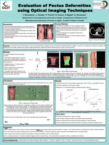
- Computed Tomography (CT) scan of patients chest
to obtain a cross sectional image of the
deformity (Figure 2). - Doctors calculate the Haller Index from the CT
scan at the point of maximum intrusion of the
deformity for Excavatum patients. - No current standard for evaluating the deformity
in Carinatum patients. - CT scans emit high levels of harmful radiation
therefore cannot be taken regularly.
- Pectus deformities are caused by an excessive
growth of the ribs and costal cartilage that
buckles inward (Excavatum) or outward (Carinatum)
(Figure 1). - The deformity affects chest wall flexibility and
hinders chest expansion during inhalation. - The deformity also affects the performance of
the heart and lungs during exercise. - Adolescent patients often have psychological
problems due to cosmetic issues.
Figure 1 Pectus Excavatum (left)
and Carinatum (right)
Figure 2 CT scan of a PE patient
Purpose The objective of the Pectus group is to
use the proposed Optical Imaging procedure
clinically at the Alberta Childrens Hospital.
In order to be accepted as a method of
evaluation, the Pectus group must obtain results
from the Optical Imaging system that correlate
well with the results from the current CT scan
procedure.
Methods
- Procedure adapted from the Scoliosis protocol.
- The Inspeck Imaging System is used to capture
and process images to create a 3-dimensional
reconstruction of the patients torso (Figure 3).
- Digitizers capture 1.3 million points of
geometry and texture. - Data is collected every month at the Alberta
Childrens Hospital. - A qualified nurse places markers on the patients
that relate internal bony structures to external
surface landmarks.
(a)
(b)
(c)
(d) (e)
(f) Figure 3 Optical Image Processing
Steps (a) Patient stands in the positioning frame
while 4 Inspeck digitizers capture images of each
side of the torso. (b) Cameras use the Moiré
Fringe Contouring technique and the Inspeck
software assigns 3D coordinates to each point on
the torso surface. (c) FAPS (Fringe Acquisition
and Processing Software) is used to select the
are of interest (torso outline). (d) Inspeck EM
application is used to merge images into a 3D
model. (e) Cross section of interest (greatest
protrusion or indentation) is located. (f) Cross
section is isolated to be further analyzed.
- Results
- 10 patients have had CTs and Optical Images
taken with HI and HImod calculated from each - 5 Carinatum patients 5 Excavatum patients
- Results from CT and Optical Imaging correlated
strongly (Figure 5)
Calculations From the optical image cross
section, the Haller Index (HI) and the modified
Haller Index (HImod) were calculated using the
steps shown in figure 4.
- (a)
(b) (c)
(d) - Figure 4 Haller Index Calculation Steps
- Cross section of maximum protrusion or
indentation is isolated. (b) Cross section is
rotated so that the line tangent to the back
humps is vertical. (c) Outermost points on the
torso are found and a transverse line is drawn
connecting them (1). Also, tangent lines are
drawn to the point of maximum deformity (2) and
spinous process (3). (d) The following
dimensions are measured dLa transverse
diameter of the chest dAP anterior-posterior
diameter at the point of greatest intrusion to
the spinous process dAPmod distance from the
point of greatest intrusion to the transverse
diameter line. - The Haller Index and modified Haller Index can be
calculated using the dimensions - HI dLa HImod dLa
- dAP dAPmod
(a)
(b) Figure 5 Correlation between (a) Haller
Indices and (b) Modified Haller Indices from
Optical Imaging and CT techniques
- Future Work
- Decrease processing time to under 30 minutes
- Create computer programs to perform calculation
steps automatically - Determine indices that can evaluate Scoliosis
progression in Pectus patients - Create a simplified format to present information
to patients and doctors (aesthetic score)
- Conclusion
- Optical Imaging method is non-invasive and can be
used regularly to monitor patient progress
through treatment or surgery. This will allow
doctors to understand the history of Pectus
disorders. - To be used clinically, the Optical Imaging
process must take under 30 minutes. Computer
programs must be produced to automatically
perform calculation steps. - Indices must me found that can evaluate the
progression of Scoliosis, that often occurs in
Pectus patients. - So far, correlations between CT results and
Optical Imaging results have been strong. More
data must be processed to further correlate the
two methods.
Acknowledgements Funding support from Markin
Flanagan USRP NSERC. D. Visser, S. Anderson,
H.Wu, K. Truong, V. Komisar from the U of C
Scoliosis Research Group.


