Biophotonics at St Andrews - PowerPoint PPT Presentation
1 / 32
Title: Biophotonics at St Andrews
1
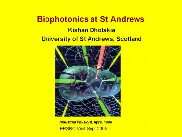
Biophotonics at St Andrews
- Kishan Dholakia
- University of St Andrews, Scotland
Industrial Physicist, April, 1999
EPSRC Visit Sept 2005
2
Biophotonics Funding at St Andrews since 1999
1999 2000 2001 2002 2003 2004
2005 2006
TOTAL 7.328M grants to KD,WS, FGM, ACR,
PEB 2006 figure is grants Jan-March (two Basic
Technology grants).
3
(No Transcript)
4
(No Transcript)
5
(No Transcript)
6
NIR short-pulse tweezing
Violet diode tweezing
NIR cw tweezing
7
Bessel beam guiding with 2-photon fluorescence
8
Using an evanescent field to manipulate particles
opens up the possibility for both enhanced
trapping near surfaces as well as the
manipulation of thousands of particles at a time.
Here we see an array of red blood cells being
trans-ported in an evanescent field above a glass
prism.
9
(No Transcript)
10
(No Transcript)
11
(No Transcript)
12
(No Transcript)
13
(No Transcript)
14
(No Transcript)
15
(No Transcript)
16
- Fluorescent proteins enable optical
- studies of cell functions.
- In this work we utilise CFP and YFP
- to quantitatively determine the binding
- affinity of proteins in vitro (as opposed
- to qualitative microscopy in vivo).
- We use fluorescence resonance energy transfer
(FRET) signal between tagged proteins as a
measure of binding - We observe a decrease in cyan and an increase in
yellow emission as energy transfer occurs at - FRET a simple and precise optical technique,
with a far more versatile range - of application than conventionally used
isothermal calorimetry.
17
A single microscope objective can be used to
deliver and capture light.
- By combining optical tweezers with a Raman
spectrometer allow us to study the evolution of
cancer in cells - Opposite are two Raman spectra of cells at
different stages in cancer evolution - This allows us to get very high sepcificity such
that we can look for differences within a
population such as might be apparent in cancer
stem cells.
18
(No Transcript)
19
(No Transcript)
20
(No Transcript)
21
(No Transcript)
22
(No Transcript)
23
Fibre trapping gives all the inherent advantages
of a fibre base approach such as ease of
delivery. It is also well suited to integration
into a microfluidic environment. The trap is
formed from two counter propagating Gaussian
beams
Can trap larger particles than any other optical
trapping approach. Can be integrated with Here we
see CHO (Chinese Hamster Ovary) cells held in a
bound array
24
(No Transcript)
25
(No Transcript)
26
(No Transcript)
27
(No Transcript)
28
(No Transcript)
29
(No Transcript)
30
(No Transcript)
31
(No Transcript)
32
Future Biophotonics Aim
- Consolidate activity
- Portfolio Grant K Dholakia, W Sibbett, F
Gunn-Moore , Andrew Riches, Paul Campbell
Explore and proliferate ideas seen as well as new
ideas to truly establish St Andrews as a place of
International bioscience


