
Chapter 16 Cell Communication - PowerPoint PPT Presentation
1 / 14
Title:
Chapter 16 Cell Communication
Description:
How cells communicate an external signal across the cell membrane. You will not be ... Pertussis toxin. Prevents GDP/GTP exchange -- GTP locked in off state ... – PowerPoint PPT presentation
Number of Views:369
Avg rating:3.0/5.0
Title: Chapter 16 Cell Communication
1
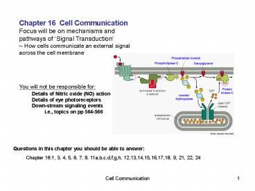
Chapter 16 Cell Communication Focus will be on
mechanisms and pathways of Signal
Transduction -- How cells communicate an
external signal across the cell membrane You
will not be responsible for Details of Nitric
oxide (NO) action Details of eye
photoreceptors Down-stream signaling
events i.e., topics on pp 564-566
Questions in this chapter you should be able to
answer Chapter 161, 3, 4, 5, 6, 7, 9,
11a,b,c,d,f,g,h, 12,13,14,15,16,17,18, 9, 21, 22,
24
2
How do cells communicate with each
other? Intracellular receptors vs
Cell-surface receptors
3
What are cellular responses to signaling
pathways? External signals trigger many
different types of cellular responses and
changes in metabolism
4
What types of molecules carry signals to
cells? 1) Steroidal hormones 2) NO (nitric
oxide) Pass into cells Bind to Intracellular
receptors 3) Peptide hormones Use cell surface
receptors
5
How do cell-surface receptors transmit signals
into the cytoplasm
An external signal activates an internal
signaling cascade
allowing amplification of a weak signal
Intracellular signaling molecules cAMP, Ca,
DAG, IP3 Downstream effectors, e.g. PKA PKC
(protein kinases) and Targets, e.g. Gene reg.
proteins metabolic enzymes
6
What are the three types of cell surface
receptors?
Ligand-gated channel
7
How are G-proteins activated? 7-pass
receptors -- Hundreds of different types --
triggering enumerable different cytoplasmic
processes Examples Glucagon activates
glucose release by liver Lutenizing Hormone (LH)
triggers progesterone release from
ovary Adrenalin (epinephrine) increases heart
rate Allergen mast cell degranulation
G-protein-linked receptors
8
How do activated G-proteins trigger release of
secondary messenger molecules? Secondary
messengers include cAMP, Ca, DAG, IP3
Some toxins interfere with G-proteins Cholera
toxin Inhibits GTPase activity of a-subunit --
causes Na efflux into intestine -- water
flow into intestine Pertussis toxin Prevents
GDP/GTP exchange -- GTP locked in off state --
mucous secretion into lungs
IP3 signaling
9
Acetylcholine acts at a G-protein-linked receptor
on heart muscle to make the heart beat more
slowly by the effect of the G protein on a K
channel, as shown in this Figure. Which one or
more of the following would enhance this effect
of acetylcholine? Explain.
(a) A high concentration of a non-hydrolyzable
analog of GTP. (b) Mutations in the
acetylcholine receptor that weaken the
interaction between the receptor and
acetylcholine. (c) Mutations in the G protein
a-subunit that speed-up the hydrolysis of
GTP. (d) Mutations in the K Channel that make
the ß?-subunit bind tighter
10
How do enzyme-linked receptors function?
Tyrosine kinases Dimerization Autophosphorylat
ion Activated signaling proteins Some
examples Cell growth factors, e.g. -- PDGF --
EGF -- trigger cell replication Insulin
triggers insulin release Antigen / antibody
B- T-cell activation
11
Signaling often occurs through Ras monomeric
GTP-binding protein Ras is an important
proto-oncogene Mutated ras can be an oncogene
12
How are complex signally pathways
dissected? Study effects of mutations and
delations in cultured cells
13
And the interactions can be complex,
indeed! Multiple signaling pathways often are
activated simultaneously
14
When activated by the signal, the
platelet-derived growth factor (PDGF) receptor
phosphorylates itself on multiple tyrosines (as
indicated below by the circled Ps the numbers
next to these Ps indicate the amino acid number
of the tyrosine). These phosphorylated tyrosines
serve as docking sites for proteins (A, B, C, and
D) that interact with the activated
PDGF-receptor. Binding of PDGF activates the
PDGF-receptor leading to an increase in DNA
synthesis. To determine whether protein A, B, C,
and/or D are responsible for activation of DNA
synthesis, you construct mutant versions of the
PDGF-receptor that retain one or more tyrosine
phosphorylation sites. In the cells, the various
versions of the PDGF-receptor become
phosphorylated on whichever tyrosines remain.
You measure the level of DNA synthesis in cells
that express the various mutant receptors and
obtain the data shown below.
- From these data, which, if any, of these proteins
A, B, C, and D are involved in the stimulation of
DNA synthesis by PDGF? Why? - Which, if any, of these proteins inhibit DNA
synthesis? Why? - Which, if any, of these proteins appear to play
no detectable role in DNA synthesis? Why? - What is the effect of the binding of A on the
effect of B?
Recommended
CrystalGraphics Presentations


