Border Molding - PowerPoint PPT Presentation
1 / 39
Title:
Border Molding
Description:
Use Bunsen Burner not Hanau Torch. Warm until it starts to droop ... To Hyoid. Mylohyoid. Ridge. X-section through. Mandibular ridge. in 2nd Molar region ... – PowerPoint PPT presentation
Number of Views:4234
Avg rating:5.0/5.0
Title: Border Molding
1
Border Molding
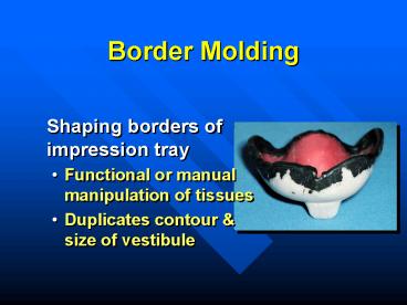
- Shaping borders of impression tray
- Functional or manual manipulation of tissues
- Duplicates contour size of vestibule
2
Border Molding
- Performed with
- Thermoplastic modeling compound
- Waxes
- Impression materials
3
Tray Wax Spacer
- Remains in place during border molding procedures
4
Custom Tray
- Comfortable
- 2-3 mm from vestibule
- Dry periphery of tray (Compound will not stick to
tray otherwise)
5
Heating Compound
- Use Bunsen Burner not Hanau Torch
- Warm until it starts to droop
- Do not overheat if catches fire or boils, it
will not mold properly
6
Compound Application
- Apply over periphery of tray, in a thickness just
slightly narrower than the compound stick
7
Re-soften After Application
- Flame with a hand torch until all seams or sharp
contours have disappeared - Do not melt wax spacer inside tray
8
Preventing Slumping
- Hold the tray upside down so that compound droops
toward the depth of the vestibule
9
Tempering Compound
- Temper in a water bath (135-140F) for several
seconds - Prevent burning
- Hot water bath will keep compound soft for an
extended period
10
Wax Spacer
- Keep out of hot water bath to prevent melting
- Difficult to replace tray intraorally in the same
position - Results in uneven border molding
11
Prepare Patient
- Patient seated, head against headrest, mouth open
relaxed - If patient opens wide, commisures constrict,
limiting access
12
Inserting the Custom Tray
- Place intraorally by rotating into place
- Mold by pulling on the cheeks, lips
- Have patient make functional movements
13
After Removal
- Chill in cold water
- Trim excess over wax spacer or external material
that is thicker than 4-5 mm - Clean debris from tray
14
Assessing Peripheral Role
- Proper thickness
- No overlap
15
Burnthrough
- Difficult to see (opaque)
- Relieve tray
16
After Trimming
- If border is sharp or has seams, re-flame, temper
and readapt intraorally - Repeat until periphery is completed
17
Border Molding
- Dont reduce border molding prior to final
impression if - Modern low viscosity materials are used
- Sufficient relief (spacer holes)
18
Maxilla - Seating the Tray
- Seat tray firmly in mid-palatal area during
border molding procedures
19
Maxilla - Contouring
- Mold posterior buccal by pulling cheek down
forward with slight circular movement
20
Functional Movements
- Patient moves mandible side to side opens wide
- Molds the retrozygomal area
- Allows for movement of coronoid process
- Prevents impingement of pterygomandibular raphe
21
Maxilla - Labial Frenum
- Pull lip outward downward
- Do not pull to one side
22
Maxilla - Labial Frenum
- Labial frenum should be narrow
- Buccal frena usually broader, V-shaped
23
Maxilla - Posterior Border
- Add compound across the top of the tray (not at
the edge)
24
Maxilla - Posterior Border
- Terminates at vibrating line and hamular notches
- Mark with an indelible stick
- Insert tray check visually
25
Evaluating Border Molding
- Relatively symmetrical
26
Evaluating Maxillary Border Molding
- Retentive
27
Mandible
- More difficult
- Changing position of the floor of the mouth
28
Posterior Buccal Areas
- Pull cheek upward while holding tray in place
- Have patient suck cheeks inward while holding
tray in place
29
Retromolar Pad
- Should be covered (at least partially) to provide
a seal and comfort to the patient
30
External Oblique Ridge
- Dont extend past EOR
- Palpate cheek at angle of the mandible
- Smooth transition between mandible border - not
palpable
31
Buccal Extension
- Look for fold in vestibule
32
Masseter Muscle
- Distal buccal extension
- Patient closes against force
- Activates the masseter, which will displace the
compound
33
Mandibular Frenal Attachments
- Labial frenum is narrow
- pull lip straight up,
- not as exaggerated as maxilla
- Buccal frena broad V-shaped
34
Posterior Lingual Areas
- Have patient touch their tongue to the corners of
the mouth, to the palate and stick their tongue
out of their mouth
35
Posterior Lingual Areas
- An S shaped lingual flange commonly results in
posterior lingual area
36
Retromylohyoid Space
- Distolingual border can extend
- Straight down from the retromolar pads
- Anteriorly to varying degrees
- Almost never angles posteriorly from retromolar
pads
37
Posterior Lingual Areas
X-section through Mandibular ridge in 2nd Molar
region
- Lower border at or slightly below mylohyoid ridge
but not deeply into the undercut below the ridge, - Minimizes, abrasion and discomfort
Buccal
Mylohyoid Ridge
Attachments To Hyoid
38
Posterior Lingual Areas
- Denture should not lift with normal tongue
movements
39
Anterior Lingual
- Patient lifts tongue to palate, to corners of
mouth and sticks tongue out - Hold tray in place denture should not lift with
normal tongue movement


