Identification of Microbial DNA in Chorioretinal Lesions - PowerPoint PPT Presentation
1 / 1
Title:
Identification of Microbial DNA in Chorioretinal Lesions
Description:
Alexandra Herzlich1, 2, Defen Shen1, Steven Yeh3, Steven M ... Fundus photo of typical choriod and retinal scars found in people with CGD. DAPI. Omi/HtrA2 ... – PowerPoint PPT presentation
Number of Views:56
Avg rating:3.0/5.0
Title: Identification of Microbial DNA in Chorioretinal Lesions
1
- Identification of Microbial DNA in Chorioretinal
Lesions - Associated with Chronic Granulomatous Disease
- Alexandra Herzlich1, 2, Defen Shen1, Steven Yeh3,
Steven M Holland3, and Chi-Chao Chan1 - 1. Laboratory of Immunology, National Eye
Institute, NIH, Bethesda, MD - 2. Department of Internal Medicine, Georgetown
University Hospital, Washington, DC - 3. Laboratory of Infectious Disease, National
Institute of Allergy and Infectious Diseases,
Bethesda, MD
PURPOSE Chronic Granulomatous Disease (CGD) is
characterized by granulomatous inflammation of
multiple systemic organs secondary to a defect in
the ability to generate reactive oxidative
species. The most common and severe form of CGD
is an X-linked recessive defect in the gene
encoding the gp91phox subunit of NADPH oxidase,
which is involved in microbial killing. This
predisposes individuals to severe, recurrent
infectious and non-infectious keratitis, and
chorioretinal lesions. We sought to identify
whether the ocular pathology associated with CGD
is caused by the same pathogens via the known
mechanisms of the systemic manifestations of this
disease.
RESULTS Of the autopsy cases of CGD-associated
deaths at the NIH from 2000 2008, six had
documented systemic manifestations from the
aforementioned organisms. Of these six, three
demonstrated chorioretinal scars associated with
CGD. P. aeruginosa and S. epidermidis DNA was
identified in two chorioretinal scars of two
different cases, whereas no evidence of other
tested microbial DNA was found. No microbial
tissue was detected in normal ocular tissue
sampled from the same eye samples.
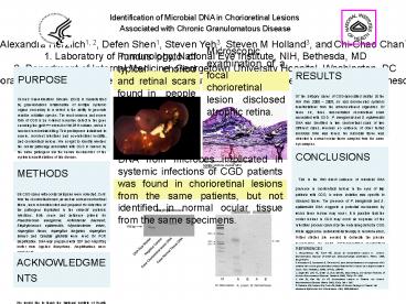
Fundus photo of typical choriod and retinal scars
found in people with CGD
Microscopic examination of a focal chorioretinal
lesion disclosed atrophic retina.
H2O2-treated
control
CONCLUSIONS This is the first direct evidence
of microbial DNA presence in chorioretinal
lesions in the eyes of twp patients with CGD, in
whom isolation was specific to diseased tissue.
The presence of P. aeruginosa and S. epidermidis
DNA suggests a potential mechanism by which these
lesions may occur. It is possible that the ocular
lesions in CGD may occur as sequelae of the
infectious process caused by the underlying
defect in CGD. While aggressive anti-microbila
therapy is recommended, further studies are
needed to delineate the precise mechanisms by
which CGD-associated chorioretinal lesions form.
Arrows Hypertrophic and hyperplastic RPE cells.
Asterisks Small area of serous retinal
detachment.
METHODS Six CGD cases with ocular autopsies were
collected. Cells from the chorioretinal scars, as
well as normal chorioretinal tissue, were
microdissected and prepared for detection of the
pathogens implicated in the subjects systemic
infections. Both sense and antisense primers for
Pseudomonas aeruginosa, Actinobacter baumanii,
Staphylococcus epidermidis, Mycobacterium avium,
Aspergillus flavus, Aspergillus funigatus,
Aspergillus terreus and Candida glabrata were
used for PCR amplification. DNA was prepared with
32P and AmpliTaq buffer from Applied Biosystems.
Amplifications were carried out.
DNA from microbes implicated in systemic
infections of CGD patients was found in
chorioretinal lesions from the same patients, but
not identified in normal ocular tissue from the
same specimens.
DAPI
Omi/HtrA2
Merged
COX IV
REFERENCES 1. Grossniklauss HE, Frank KE, Jacobs
G. Chorioretinal Lesions in Chronic
Granulomatous Disease of Childhood.
Clinicopathologic Correlations. Retina. 1988
8(1)270-274. 2. Kobayashi S, Murayama S,
Takanashi S, et.al. Clinical features and
prognoses of 23 patients with chronic
granulomatous disease followed for 21 years by a
single hospital in Japan. Eur J Pediatrics. 2008
167(12)1389-94. 3. Segal BH, Leto TL, Gallin
JI, et.al. Genetic, Biochemical, and Clinical
Features of Chronic Granulomatous Disease.
Medicine. 2000 79(3)170 200. 4. Kim SJ, Kim
JG, Yu YS. Chorioretinal Lesions in Patients with
Chronic Granulomatous Disease. Retina. 2003
23(3) 360 365. 5. Palestine AG, Meyers SM,
Fauci AS, Gallin JI. Ocular Fiindings in Patients
with Neutrophil Dysfunction.Am J Ophthalmol.
1983 95(5)598-604.
DAPI
Omi/HtrA2
DAPI
Omi/HtrA2
ACKNOWLEDGMENTS We would like to thank the
National Institute of Health Intramural Research
Program for their support in the funding of this
project.
Merged
COX IV
COX IV
Merged


