Problems in Twin Pregnancy - PowerPoint PPT Presentation
1 / 2
Title:
Problems in Twin Pregnancy
Description:
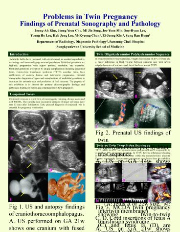
Fig 1. US and autopsy findings of craniothoracoomphalopagus. ... G.Autopsy specimen of another acardiac twins shows absent head, thorax and upper ... – PowerPoint PPT presentation
Number of Views:847
Avg rating:3.0/5.0
Title: Problems in Twin Pregnancy
1
Problems in Twin Pregnancy Findings of Prenatal
Sonography and Pathology Jeong-Ah Kim, Jeong Yeon
Cho, Mi Jin Song, Jee-Yeon Min, Soo-Hyun
Lee, Young Ho Lee, Hak Jong Lee, Yi-Kyeong Chun1,
Ei-Jeong Kim1, Sung Ran Hong1 Department of
Radiology, Diagnostic Pathology1, Samsung Cheil
Hospital Sungkyunkwan University School of
Medicine
Twin Oligohydramnios Polyhydramnios Sequence
Introduction
Multiple births have increased with development
in assisted reproductive technology and increased
aging maternal population. Multifetal gestations
are high-risk pregnancies with higher perinatal
morbidity and mortality. Multifetal gestations
are subject to unique complications including
conjoined twins, twin-to-twin transfusion
syndrome (TTTS), acardiac twins, twin emblization
of co-twin demise and heterotopic pregnancies.
Prenatal sonographic diagnosis of types and
complications of multefetal gestations is
important for antenatal care and prediction of
fetal outcome. The purpose of this exhibition is
to present the prenatal ultrasonographic findings
and pathologic findings of the unique
complicaitons of twin pregnancy.
In monochorionic twin pregnancies, weight
discordance of 20 or more and a major difference
in fluid volume between amniotic sacs with severe
oligohydramnios of one sac (stuck twin) has been
termed TOPS.
Conjoined Twins
Conjoined twins are a rarest form of monozygotic
twinning, always associated with MCMA. They
results from incomplete division of inncer cell
mass more than 13 days after fertilization. Early
prenatal diagnosis of conjoined twin is essential
for pregnancy termination .
Fig 2. Prenatal US findings of twin
oligohydramnios/polyhydramnios sequence. A. US on
GA 14w shows single placenta with intertwin
membrane (arrow). B. US on GA 22w shows stuck
twin (B, arrow) of 20w size with severe
oligohydramnios. (A fetus A of 22w size,
M intertwin membrane) C, D. Cord insertions
of fetus A (C) and fetus B (D) are separated
without anastomosis.
Twin-to-Twin Transfusion Syndrome
TTTS occurs in 4-35 of monochorionic
pregnancies and is responsible for as much as 17
of prenatal mortality. In TTTS, there is
unbalanced shunting in the deep arteriovenous
anastomoses within the monochorionic placenta.
Doppler sonography demonstrates antepartum
transfusion from the umbilical artery of donor
twin into the umbilical vein of the recipient.
Fig 1. US and autopsy findings of
craniothoracoomphalopagus. A. US performed on GA
21w shows one cranium with fused cerebrum and one
cerebellum. B. Fused anterior chest with fused
two hearts (H). C. Fused cranium with V shaped
two spines (arrows). D. Fused anterior abdomen
with two ischiums (ISC). E.F.Specimen
radiographs(E) and autopsy (F) show a
craniothoracoomphalopagus.
Fig 3. MCDA twin pregnancy showing twin-to-twin
transfusion syndrome. A. US on GA 21w shows
smaller fetus with oligohydramnios (B) and larger
one (A). B. Significant discrepancy of fetal
sizes. Fetus A is larger than fetus B by more
than 2 SD. C, F. CDUS shows approximate insertion
of umbilical cords D, E. Umbilical arterial
duplex US of smaller fetus (D) shows increased
vascular resistance and that of larger fetus (E)
shows normal flow pattern.
2
Acardiac Twins
Co-Twin Demise
The incidence of spontaneous single intrauterine
loss is 2.6-6.8 . The complications of single
fetal demise are preeclampsia, preterm labor and
intrauterine growth retardation. In monochorionic
pregnancies, increased risk of cerebral necrotic
lesion due to thromboemblic material, anemia in a
twin transfusion syndrome or premature delivery
have been reported.
Acardia is a lethal anomaly occurring in 1 of
monozygotic twin. Acardia develops during early
embryogenesis as a result of extensive
artery-to-artery and vein-to-vein anastomosis
through monochorionic placenta with reverse
umbilical arterial circulation. The acardiac twin
has a parasitic existence and depends on the
donor (pump) twin for its blood supply via
placental anastomoses and retrograde perfusion of
umbilical cord. Perfusion of malformed fetus
occurs via artery-to-artery anastomosis between
the fetuses. This twin reversed arterial
perfusion (TRAP) sequence is a most extreme
manifestation of the TTTS. Depending on the state
of disruption, acardiac anomalies are divided
into four categories acardius anceps, acardius
acephalus, acardius acormus and acrdius amorphus.
Doppler verification of reversed flow in
umbilical cord of the acardiac twin confirms the
diagnosis.
A
B
Fig.5. Sonographic findings of MCDA twins with
co-twin demise A. US performed on GA 14w shows
thin intertwin membrane (arrow) with single
placenta and IUFD of one fetus with diffuse soft
tissue edema (double head arrow). B. US on GA 23w
shows cystic hygroma of the dead fetus. C.
Enlarged umbilical cord was noted in the
surviving fetus in the US on 23w. D.
Cardiomegaly with thickened biventricular walls
in surviving fetus.
Heterotopic Pregnancy
D
C
Treatment for ovulation induction and assisted
reproductive technology have dramatically
increased the incidence of heterotopic pregnancy.
The incidence of heterotopic pregnancy is 1 in
ovulation induction procedure.Sonographic
detection of an extrauterine gestational sac with
or without a fetal pole with an IUP confirm the
diagnosis. Most frequent location is tube
(ampulla gtisthmic gtfimbria). The prognosis of IUP
is similar to that without heterotopia.
AO
E
F
Fig.6. Tubal heterotopic pregnancy. A. US on GA
8w shows fetal pole (calipers) within the
intrauterine gestational sac (arrow). B.
Simultaneous extrauterine gestational sac (arrow)
located between uterus (UT) and left ovary
(LO). C. Left salpingectomy verified ectopic
pregnancy demonstrating the hemorrhagic mass
(arrow) within the dilatated left fallopian tube.
H
G
Fig.4. Sonographic diagnosis and autopsy findings
of acardiac twins in monochorionic pregnancy. A.
US performed on GA 19w shows significant
discrepancy of fetal sizes. Absence of heart and
diffuse soft tissue edema are demonstrated in the
larger fetus B. B. No head in fetus B(arrowhead)
with rudimentary bony upper extremities
(arrow). C. Multiseptated cystic mass suggesting
large cystic hygroma is noted along back of
fetus. D. CDUS demonstrates interfetal
anastomoses of umbilical veins and arteries.
Fetus B have singe umbilical artery and vein. E.
Duplex US verify the reversed flow in umbilical
artery (UA) of acardiac fetus B. F. Reversed flow
downside up is noted in descending aorta (AO) of
acardiac fetus B. G.Autopsy specimen of another
acardiac twins shows absent head, thorax and
upper extremities with knotting of umbilical cord
(arrow). H. Monochorionic placental tissue shows
artery-to-arterial anastomosis of umbilical
vessels including single umbilical arteries of
both fetuses.
Fig.7. Cornual heterotopic pregnancy. A.
Gestational sac is seen within the uterine cavity
(arrow). B. Another gestational sac (yellow
arrow) with embryo (red arrow) in the cornual
portion of uterus.


