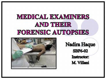
MEDICAL EXAMINERS AND THEIR FORENSIC AUTOPSIES - PowerPoint PPT Presentation
1 / 27
Title: MEDICAL EXAMINERS AND THEIR FORENSIC AUTOPSIES
1
MEDICAL EXAMINERS AND THEIR FORENSIC AUTOPSIES
- Nadira Haque
- SBF6-02
- Instructor
- M. Villani
2
What is a Medical Examiner?
- A medical examiner is a doctor specialized in
- anatomical pathology
- forensic pathology
- The Medical Examiner performs autopsies on bodies
to determine - How, When, and Where the victim died.
3
Coroner Versus Medical Examiner
Nadira Haque SB5-02 M. Villani
- Coroners, unlike Medical Examiners, are not
Medical Doctors. - Coroners provide subjective opinions of the
situation. - Medical examiners provide objective opinions
based on their observations.
4
Qualifications of a Medical Examiner
Nadira Haque SB5-02 M. Villani
- After high school, education includes
- 4 years undergraduate training
- 4 years medical school
- 4 years residency in anatomic and clinical
pathology - 1 year pathology fellowship
- MEs must pass Medical Boards in both topics
- In order to maintain a license, 20 hours per year
of approved medical education is necessary.
5
The Forensic Autopsy
Nadira Haque SB5-02 M. Villani
- Forensic pathology was first recognized in the
United States in 1959. - A forensic autopsy is an examination of a corpse.
This is done to determine the - cause and manner of death
- time of death
- location of death
- The body is examined internally and externally.
6
The Important Contents of an Autopsy Report
Nadira Haque SB5-02 M. Villani
- Autopsy reports contain objective opinions from a
medical examiner - These report includes the
- The Cause of Death based on the Autopsy
- The circumstances surrounding the cause of death
- All wounds and injuries found on body
- A report on the bodily fluids and its contents
7
Circumstances of Death
Nadira Haque SB5-02 M. Villani
- The circumstances of a death refers to the
manner of death. - The major categories are
- To pathologists, the subcategories of unnatural
death include
8
Determining Time of Death Algor Mortis
Nadira Haque SB5-02 M. Villani
- Algor Mortis is known as the coolness of death
- Normal body temperature is 98.6o F.
- Body temperature decreases 2o C in the first hour
if in a controlled environment - Then the body temperature is decreased 1o C per
hour, after the first hour. - This occurs due to the respiratory system failing
to work after death
9
Time of Death Livor Mortis
- Livor Mortis is referred to as color of death.
- When the heart stops pumping, blood no longer
flows through the body. Therefore, red blood
cells gather with plasma cells at the lowest
point of the body. - If the body is not moved, there will be a visible
area of coloration.
10
Time of Death Rigor Mortis
- Rigor Mortis is known as stiffness of death.
- A dead body stiffens in this order eyelids, jaw,
face, arms, legs, torso. - Rigor mortis shows up after 2 hours of death
- Body stiffness peaks at 12 hours
- At 12-24 hours, the body begins loosing stiffness
- After 24 hours, rigor mortis is no longer present
11
Time of Death Pallor Mortis
- Pallor Mortis is referred to as paleness of
death. - This identification is only useful if the body is
found quickly after it has died. - Capillary circulation stops when a person dies,
thus giving the paleness - It shows up 15-20 minutes after death.
12
Protocol for External Examination
Nadira Haque SB5-02 M. Villani
- At the scene, the body goes into a body bag.
- The body is then brought into the medical
examiners room where it is weighed. - It is then placed on a cadaver dissection table.
- Gender, race, hair, age and other defining
factors are noted of.
13
Wounds found in External Examination of Body
Nadira Haque SB5-02 M. Villani
- Wounds are noted of and photographed.
- Common injuries include
- Bruises (contusions) black and blue marks.
- Abrasions made by dragging and scraping.
- Lacerations ragged cuts
- Cuts superficial injuries
- Marks of ligature indicating torture and being
bound - Pinpoint hemorrhages
14
Internal Examination of Body Autopsy Process (1)
- Step 1 A body block made of plastic or rubber
is used. It props up the back, and allows the
arms and head move backwards. - Step 2 A deep Y-shaped cut is made, starting
from the armpits, down the front of the chest,
and ending at the sternum. The incision is then
extended to the pubic bone. The cuts are made to
the left.
15
Internal Examination of Body Autopsy Process (2)
- Step 3 An electric saw is used to cut open the
chest. This creates dust, so the cavity is opened
using Shears. - Step 4 The ribs are cut to make the sternum and
ribs one removable piece. This exposes the lungs
and heart. The chest plate is put aside.
16
Internal Examination of Body Autopsy Process (3)
- Step 5 The organs are then removed.
- The en masse technique of letulle Organs are
removed as one large mass. - Cuts are made along the vertebrae Organs are
detached. - The en bloc method of Ghon Organs divided into
four sections and removed as four sections
17
Internal Examination of Body Autopsy Process (4)
- Step 6 The organs are examined and weighed.
Tissue samples from each organ are taken. - Step 7 The major blood vessels are cut open and
inspected for any unnatural blocks or content. - Step 8 The stomach and intestinal contents are
examined and weighed.
18
Internal Examination of Body Autopsy Process (5)
- Step 9 The body block is shifted, to prop up the
head. Now, the skull is opened. - An incision is made from one ear, over the crown,
to the other ear. - The scalp is pulled away in two flaps
- A cap of the skull is cut using an electric
saw the brain is now exposed. - The brain is observed for any abnormalities or
damage.
19
Internal Examination of Body Autopsy Process (6)
- Step 10 The autopsy is finished. The body is now
reconstructed. This process is usually assisted
with filler material, such as cotton. - Organs are put into plastic bags before being put
back into the body cavity in order to prevent
leakage. - All open areas are fitted back and sewn/sealed so
the body can be returned to the family.
20
The Lungs
Nadira Haque SB5-02 M. Villani
- In humans, the right lung is larger than the
left. - Pink lungs belong to younger people.
- With age, the lungs turn a grey color due to the
amount of gas inhaled throughout a persons life.
- The alveoli are closed in the lungs due to the
capillaries not receiving a gas exchange - If there is soot and grime in the lungs, the
victim may have died in a fire. - Water found in lungs indicates drowning.
21
The Kidneys
Nadira Haque SB5-02 M. Villani
- The left kidney is larger than the right.
- As a person gets older
- Their kidneys shrink and become more wrinkly
- These traits in a kidney indicate disease
- Extreme sponginess.
- Presence of cysts.
22
The Liver
Nadira Haque SB5-02 M. Villani
- The weight of the liver in a adult is 3 pounds.
- A normal liver is reddish-brown in color.
- A pale-yellow and fatty liver indicates a heavy
drinker, which could be the natural cause of
death.
23
Stomach Contents
Nadira Haque SB5-02 M. Villani
- Stomach contents can be useful in determining the
time of death. - Food naturally passes through the bowels during
digestion. Digestion occurs within 2 hours of
consumption. - If large fragments of food are still present in
the stomach, the person most likely died not too
long after he or she ate. - The emptier the stomach content area, the longer
the victim has been dead.
24
Collection of Body Fluids
Nadira Haque SB5-02 M. Villani
- Body fluids are collected with syringes.
- Blood is collected from the left ventricle of
heart - It Contains DNA for identification.
- Urine is collected from the kidneys where the
nephrons are located - Urine can show the presence of toxic substances.
- Spinal fluid is collected from a canal at the
center of the spine through a spinal tap. - Joint fluid is collected from ball and socket
joins. - Vitreous humor fluid is collected from the eye
- The more potassium that has been built up, the
longer the person has been dead.
25
Toxicological Analyses
Nadira Haque SB5-02 M. Villani
- Fluid samples are sent to the toxicology division
for qualitative and quantitative analysis. - Gas chromatography provides a chart that
identifies all components in the fluid and how
much of each component is in the fluid. - Sometimes poisons, like cyanide, or drugs, such
as heroin, is the cause of death.
26
Testifying in Court
Nadira Haque SB5-02 M. Villani
- Medical examiners serve as expert witnesses in
the courtroom. - They provide the circumstances surrounding the
death based on their medical findings. - The State Medical Examiner must clear during his
or her testimony and present the medical
information in a way that the jury can
understand.
27
References
- Info http//www.forensicmed.com/faq_forensicpath.
htm - Image http//blog.nj.com/iamnj/2007/09/large_hua1
.jpg - Info http//en.wikipedia.org/wiki/Coroner
- Image http//www.theage.com.au/ffximage/2008/01/2
3/body_wideweb__470x308,0.jpg - Info http//en.wikipedia.org/wiki/Forensic_pathol
ogy - Image http//www.yogacoffeeoutlook.com/autopsy.jp
g - Info http//www.maricopa.gov/Medex/faq.aspx
- Imagehttp//www.trotman.co.uk/files/books/large/2
20508102137--Medical20School20WEB.jpg - Info http//en.wikipedia.org/wiki/Coroner
- Image http//toppayingideas.com/blog/wp-content/u
ploads/2008/04/the-homicide-report.jpg - Info http//www.hbo.com/autopsy/baden/qa_4.html
- Image http//www.homicidetraining.com/images/inse
rt.jpg - Info http//www.hbo.com/autopsy/baden/qa_4.html
- Images http//americasunknownchild.net/l
- Info http//www.deathonline.net/decomposition/for
ensic/timing.htm - Image http//www.exploreforensics.co.uk/images/11
778.jpg - Info http//en.wikipedia.org/wiki/Pallor_mortis
- Imagehttp//www.geradts.com/anil/ij/vol_004_no_00
2/papers/paper001/1.jpg - Infohttp//www.uchsc.edu/pathology/department/doc
uments/ForensicAutopsyProtocol.pdf
- Info http//www.pathguy.com/autopsy.htm
- Imagehttp//www.startsmartads.com/images/game2_au
topsy.jpg - Info http//www.pathguy.com/autopsy.htm
- Imagehttp//cidc.library.cornell.edu/dof/italy/im
ages/Musso/Autopsy.jpg - Infohttp//www.deathonline.net/what_happens/autop
sy/autopsy_steps.cfm - Imagehttp//upload.wikimedia.org/wikipedia/common
s/thumb/f/fd/Streptococcus_pneumoniae_meningitis,_
gross_pathology_33_lores.jpg/402px-Streptococcus_p
neumoniae_meningitis,_gross_pathology_33_lores.jpg
- Infohttp//www.deathonline.net/what_happens/autop
sy/autopsy_steps.cfm - Imagehttp//tbn1.google.com/images?qtbnYhh1Oyg1
HDkiLMhttp//www.charonboat.com/2007/10/charonboa
t_dot_com_woman_after_autopsy_1.jpg - Info http//www.pathguy.com/autopsy.htm
- Imagehttp//oac.med.jhmi.edu/cpc/images/cpc2/03.j
pg - Infohttp//www.pubmedcentral.nih.gov/articlerende
r.fcgi?artid1863412 - Imagehttp//www.nlm.nih.gov/visibleproofs//media/
large/v_a_408_autopsy.jpg - Infohttp//www.pubmedcentral.nih.gov/articlerende
r.fcgi?artid1863412 - Imagehttp//www.nlm.nih.gov/visibleproofs//media/
large/v_a_408_autopsy.jpg - Info http//www.pathguy.com/autopsy.htm
- Image http//scotthull.files.wordpress.com/2007/0
1/scotts-liver-cropped.JPG - Infohttp//www.exploreforensics.co.uk/stomach-con
tents-as-a-means-of-evidence.html - Imagehttp//www.fisheries.org/units/education/fis
heries_techniques/Chapter17/Subsampling20zooplank
ton20from20the20stomach20contents20of20a20f
ish.jpg - Infohttp//www.vifm.org/fp_autopsyprocess.html

