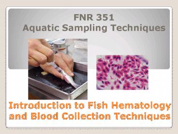
Introduction to Fish Hematology and Blood Collection Techniques - PowerPoint PPT Presentation
1 / 27
Title:
Introduction to Fish Hematology and Blood Collection Techniques
Description:
Introduction to Fish Hematology. and Blood Collection Techniques. FNR 351 ... Familiarize with most the important techniques to evaluate red and white blood ... – PowerPoint PPT presentation
Number of Views:4304
Avg rating:3.0/5.0
Title: Introduction to Fish Hematology and Blood Collection Techniques
1
Introduction to Fish Hematology and Blood
Collection Techniques
- FNR 351Aquatic Sampling Techniques
2
Objectives
- Understand the importance of blood sampling in
fishes - Introduce concepts related to hematopoiesis
- Familiarize with most the important techniques to
evaluate red and white blood cell status in
fishes - Understand basic interpretation of these values
and what they mean
3
Why is blood sampling important?
- ________________
- ________________
- ________________
- ________________
4
Common sources of variation and error
- Many intrinsic and extrinsic factors are known to
affect blood parameters in fish - Usually, the combination of ______________________
__________________________________________________
_______________
5
Can we establish Normal hematological values in
fish?
- Fish are __________________
- Fish are always in an adapting state
- So can we talk about ________ hematological
values in fish?
6
Hematopoiesis
- Definition of hematopoiesis
- Fish hematopoietic tissues
- ____________
- ____________
- ____________
7
Hematopoiesis
- Fish hematopoietic tissues
- Thymus
- Location and structure vary with species
- ___________________________
- Most cells found are lymphocytes
8
Hematopoiesis
- Fish hematopoietic tissues
- Spleen
- Usually found _____________________________
- Contains _____ and ______ pulp areas
- Contain cells involved in hematopoiesis
(production of red blood cells, white blood cells
and trombocytes)
9
Hematopoiesis
- Fish hematopoietic tissues
- Kidney
- Major blood-forming organ of bony fish
- Can de divided in two components
- Cranial kidney ? head kidney
- Site of _______________(erythropoiesis)
- Site of _______________(lymphopoiesis)
- Also involved in the production of antibodies
10
Hematopoiesis
11
Hematopoiesis
Heterophils
12
Hematopoiesis
Thrombocyte
Erythrocyte
Neutrophil
HEMOCYTOBLAST
Monocyte
Plasma Cell
Lymphocyte
Macrophage
13
Hematopoiesis
- Red Blood Cells
14
Hematopoiesis
- White Blood Cells
15
Hematopoiesis
16
Techniques Blood Collection
- Several techniques are available, depending on
fish size and whether fish want to be maintained
alive or not - Small fishes (lt 8 cm TL), terminal
- Large fishes, non-terminal
- Large or Small, non-terminal
17
Techniques Blood Collection
18
Techniques Red Blood Cells
- RBC (or erythrocyte) status is usually described
in terms of the following parameters - a) Hemoglobin content (Hb)
- b) Hematocrit (Hct) or Packed Cell Volume (PCV)
- c) Red Blood Cell counts (RBC)
- d) Red Blood Cell Morphology
19
Techniques Red Blood Cells
- 1) Hemoglobin (Hb)
- Indicator of ____________________
- ______________________________
- Method of choice ______________________________
- Whole blood (20 µL) is mixed with cyanide
reagents (e.g. KCN) and the amount of
cyanomethemoglogin (Hb-CN-) measured - Units g/100 mL whole blood
20
Techniques Red Blood Cells
- 2) Packed Cell Volume or Hematocrit
- Simple and rapid test used to evaluate the amount
of RBCs - Samples are centrifuging for 5-10 min in a
microhematocrit centrifuge at 7,000 RPM - Hematocrit is calculated as _________
- _______________________________
- Results are expressed as
21
Techniques Red Blood Cells
- 3) RBC Numbers cells/µL of whole blood
Hemocytometer
22
Techniques Red Blood Cells
- Hb, RBC numbers, and PCV are used to calculate
the following indices - Mean Corpuscular Volume (MCV)
- MCV PCV () x 10 reported in nm3
- RBC (106 mm3)
- Mean Corpuscular Hemoglobin (MCH)
- MEH __Hb (g/L) reported in µg/cell
- RBC (106 mm3)
- Mean Corpuscular Hemoglobin Concentration (MCHC)
- MEHC Hb (g/100 mL) reported in g/100 mL
- PCV ()
23
Techniques Red Blood Cells
- 4) Red Blood Cell Morphology Blood Smears
- Used to evaluate __________________and cell
_____________________in peripheral blood - Also used to examine for the presence of
________________ and ______________ - Thin smears consist of blood spread in a layer
such that the thickness decreases progressively
toward the feathered edge - Slides are first air dried fixed in methanol for
5 min washed with distilled water stained
(Giemsa, Wrights, etc.) for 10-20 min washed
again examined under light microscope (100X with
immersion oil)
24
Hemolymph Smear
Bitter Crab Syndrome
Source www.afsc.noaa.gov/.../jfm04/images/RACE_fi
g4.jpg
A
B
Cells from hemolymph smears stained with Giemsa
stain modified (Sigma) at 250X magnification. A.
Crab hemocytes. B. Hematodinium sp (center
cell).
25
Techniques White Blood Cells
- 1) WBC Counts
- Similar method as described earlier for RBC
counts - 2) Differential WBC Counts
- Express the percentage distribution of each WBC
type to the total WBC population - This is done by staining blood smears and
examining them under the microscope
26
Interpretation of Values RBC
- Decrease in RBC numbers, sizes, and/or Hb content
? __________ - Increase in RBC numbers ?
- Real increase is called ________________(immatu
re stages abundant) - Artifact due to dehydration (cell morphology
normal)
27
Interpretation of Values WBC
- Decrease in WBC numbers (leukopenia) ?
- Lymphopenia
- Heteropenia
- Increase in WBC numbers (leukocytosis) ?
- Heterophilia
- Lymphocytosis


