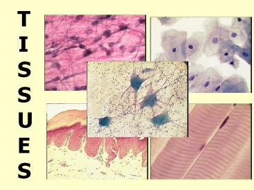
TISSUES - PowerPoint PPT Presentation
1 / 79
Title: TISSUES
1
TISSUES
2
I. Introduction
- Basics
- Tissues
- Group of cells with similar structure and
function - 2. The study of tissues
- Histology
3
I. Introduction
- Basics
- 3.Tissues consist of two
components - a) cells
- b) extracellular matrix of
- water
- small solutes
- fibrous proteins
4
I. Introduction
- There are 4 tissue types
- 1. Epithelial tissue
- protection
- 2. Connective tissue
- support
- 3. Muscle tissue
- movement
- 4. Neural tissue
- control
5
(No Transcript)
6
(No Transcript)
7
II. Epithelial Tissue
- A. Location
- includes outer layer of skin
- lines the tubes and open cavities of the
digestive and respiratory systems - lines blood vessels heart
8
II. Epithelial Tissue
- A. Location
- covers walls organs of ventral body cavity
- forms glands sense organs
9
II. Epithelial Tissue
- B. Functions
- 1. Protects from mechanical chemical injury
- 2. Protects against bacterial invasion
- 3. Filters, absorbs, secretes
10
II. Epithelial Tissue
- C. Description
- tightly packed cells
- little or not visible matrix
- may have singular or multiple layers
11
C. Description
- has a free surface
- is firmly attatched to a basement membrane
- 6. can divide rapidly (regeneration)
12
C. Description
- is avascular
- receive nutrients by diffusion
13
D. Classification
- 1. Number of cell layers
- a. Simple
- Function absorption, secretion, filtration
- single layer of cells each cell extends from
the basement membrane to the free surface.
14
D. Classification
- 1. Number of cell layers
- b. Stratified
- more than one layer of cells
- only one layer is adjacent to the basement
membrane. - high stress areas
15
D. Classification
- 1. Number of cell layers
- c. Pseudostratified
- 1 layer of cells
- Each cell touches basement membrane
- Appears stratified
16
II. Epithelium
- D. Classification
- 1. Number of cells
- 2. Cell Shape
- a. Squamous
- Squashed, scale like
- Flat nucleus
17
2. Cell Shape b. Cuboidal
- Squarish or hexagonal in profile
- Nucleus round centrally located
18
D. Classification
- 1. Layers
- Cell Shape a. Squamous
- b. Cuboidal c. Columnar
- longer than wide
- cylindrical
- nucleus near basement membrane
19
D. Classification
- 2. Cell Shape
- d. Transitional
- combination of shapes
- tolerates stretching
20
(No Transcript)
21
II. Epithelial Tissue
- E. Endothelium
- 1. Covers and lines
- a) heart
- b) blood vessels
- 2. Simple squamous
- slick thin
- capillaries are permeable
22
II. Epithelial Tissue
- F. Epithelial Membranes
- 1. Epithelium connective tissue small
organs - 2. Mucosae membranes
- Line organs, body cavities
- Open to exterior
- Respiratory, digestive urogenital
23
F. Epithelial Membranes
- 2. Mucosae membranes
- Lining of mouth stratified squamous
- Lg Sm Intestine columnar
24
F. Epithelial Membranes
- 3. Serosa
- Lines ventral, visceral, parietal cavities
walls of air sacs in lungs simple squamous
25
F. Epithelial Membranes
- 3. Synovial
- Lines joints
- cushions, protects
26
G. Cutaneous Epithelium
- 1. Is the skin
- Keratinized, dry epidermis
- 2. Epidermis is stratified squamous
27
II. Epithelium
- H. Glandular Epithelium
- 1. Characteristics
- a) cells specialized to produce and
secrete substances - b) cells typically columnar or cuboidal
- c) Kinds
- Endocrine Exocrine
28
(No Transcript)
29
H. Glandular Epithelium
- 2. Endocrine
- a) produce hormones which
- regulate or coordinate other cells
30
H. Glandular Epithelium
- 2. Endocrine
- b) ductless
- release secretions into the blood
31
H. Glandular Epithelium
- 2. Endocrine
- c) Examples thyroid, pituitary,
adrenal
32
THYROID GLAND
LUMEN
33
H. Glandular Epithelium
- 3. Exocrine
- a) release secretions into the into ducts
- b) Examples salivary, mammary,
- sweat,
- oil
34
E. Glandular Epithelium
- 3. Exocrine
- c) Structure
- simple - unbranched
- compound branched
35
E. Glandular Epithelium
- 3. Exocrine
- c) Structure
- tubular uniform diameter
36
E. Glandular Epithelium
- 3. Exocrine
- c) Structure
- acinar secretory cells in sac at
end
37
E. Glandular Epithelium
- 3. Exocrine
- c) Structure
- tubuloacinar
- secretory cells in both sac and
tube
38
(No Transcript)
39
Connective TissueThe most abundant tissue
- A. Functions
- bind structures together
- tendons
- provide support
- bones
- protection
- cartilage
- insulation
- fat
- transportation
- blood
40
III. Connective Tissue
- B. Description
- cells scattered among fibers and matrix
- intercellular material
- Vascular to avascular
- Cells capable of division
41
III. Connective Tissue
- C. Kinds of Fibers (Protein)
- Collagen
- Strong flexible
- Reticular
- Fine branching fibers form supporting framework
- Elastic
- stong elastic
42
III. Connective Tissue
- D. Kinds of Cells
- Fibroblast
- Makes fibers matrix
- Fibrocyte
- Mature fibroblast
- 3. Macrophage
- Defense, phagocytosis
- Plasma cell
- Makes antibodies
43
III. Connective Tissue
- D. Kinds of Cells
- 5. Mast Cells
- Release heparin (anti-coagulant)
- Release histamine (dilates small blood vessels)
- 6. Fat Cells
- Mature fibroblast
44
(No Transcript)
45
III. Connective Tissue
- E. Areolar connective tissue 1. Structure
- collagenous elastic fibers
- all 6 types of connective tissue cells
- filmy matrix
46
III. Connective Tissue
- E. Areolar connective tissue 2. Function
- Covers organs
- Holds vessels nerves in place
- 2nd line of defense
- Location
- Mucous membranes
- Under skin
- Between tissues of organs
47
III. Connective Tissue
- F. Dense connective tissue
- Structure
- Collagen fibers
- Fibroblasts and macrophages
- Dense matrix
48
III. Connective Tissue
- F. Dense connective tissue
- Function
- Provide strength
- Location
- Tendons
- Ligaments
49
III. Connective Tissue
- G. Elastic connective tissue
- Structure
- elastic fibers
- Few fibroblasts
- Function
- Allows stretching
- Location
- Lungs
- Trachea
- Arteries
50
III. Connective Tissue
- H. Reticular connective tissue
- Structure
- Reticular fibers
- Thin matrix
- Function
- Hold cells of loose organs together
- Location
- Liver
- Spleen
- Bone marrow
51
III. Connective Tissue
- I. Adipose connective tissue
- Structure
- collagenous elastic fibers
- all 6 types of connective tissue cells
- Fibroblasts specialize in fat production
52
III. Connective Tissue
- I. Adipose connective tissue
- Function
- Food reserve
- Prevent loss of body heat
- Location
- Around most organs
- Beneath skin
- Marrow of long bones
53
III. Connective Tissue
- J. Cartilage
- General Characteristics
- Chondrocytes
- matrix is semisolid to solid gel
- lacuna - a depression in matrix which houses
chondrocytes - perichondrium - connective tissue membrane around
cartilage - no blood supply
54
III. Connective Tissue
- J. Cartilage
- Kinds
- Hyaline
- Tip of nose
- Sternum
- Growth plate of bones
- Developing embryo
Stiff but flexible
55
III. Connective Tissue
- J. Cartilage
- Kinds
- Fibrocartilage very tough
- Between vertebrae
- Pubic symphysis
- Elastic cartilage very flexible
- External ear
- Larynx
- Eustachian tubes
56
III. Connective Tissue
- K. Bone
- General Characteristics
- organic matter - 35 (cells
fibers)
- inorganic material - 65
- (matrix
- calcium salts)
57
III. Connective Tissue
- K. Bone
- 2. Types of bone cells
- osteocytes - maintenance of matrix
- osteoblasts - bone forming cells
- osteoclasts - destroy bone matrix
58
III. Connective Tissue
- K. Bone
- 3. arranged into concentric rings called
Haversian systems - lamella - concentric ring of matrix
59
III. Connective Tissue
- K. Bone
- 3. arranged into concentric rings called
Haversian systems - lacuna - openings between lamellae for osteocytes
60
III. Connective Tissue
- K. Bone
- 3. arranged into concentric rings called
Haversian systems - osteocytes - mature bone cell
61
III. Connective Tissue
- K. Bone
- 3. arranged into concentric rings called
Haversian systems - Haversian canal - in center of lamella houses
vessels
62
(No Transcript)
63
III. Connective Tissue
- J. Bone
- 3. arranged into concentric rings called
Haversian systems - Canaliculi - radiating channels between lacuna
and Haversian canal for nutrients and wastes
64
(No Transcript)
65
III. Connective Tissue
- K. Bone
- 3. arranged into concentric rings called
Haversian systems - Volkmann canal - crosswise canals from Haversian
canal to exterior containing blood vessels
and nerves
66
IV. Connective Tissue
- L. Blood
- Characteristics
- Only fluid tissue
- Fluid matrix called plasma
- Dissolved fibers in plasma
- Cells
67
- Cells
- erythrocytes (RBC) carries O2 CO2
- leukocytes (WBC)
- immunity, defense
- thrombocytes (platelets) blood clotting
- lymph takes interstitial fluid back to blood,
immunity
68
(No Transcript)
69
IV. Connective Tissue
- L. Blood
- Location
- within blood vessels
- Function
- transport of gases, nutrients, wastes
70
V. Muscle Tissue
- A. Contractile Tissue Produces
- Movement
- Heat
B. 3 Kinds
71
V. Muscle Tissue
B. 3 Kinds
- Skeletal
- Striated
- Voluntary
- multiple nuclei
72
V. Muscle Tissue
B. 3 Kinds
- 2. Smooth
- non striated
- Involuntary
- spindle shaped
- walls of visceral organs
73
B. 3 Kinds
- 3. Cardiac
- Striated
- Involuntary
- Branching cells
- intercalated discs between cells
- heart
- can't divide or replace cells if injured
74
VI. Neural Tissue
A. Function
- conduction of information or instructions in the
form of electrical impulses
75
VI. Neural Tissue
B. 2 Kinds of Cells
- neurons
- Conducting cells
- can't divide,
- 100,000 brain cells lost daily!
- neuroglia
- support, nourish neurons
- limited repair of axons
76
VI. Neural Tissue
C. Typical Neuron
- dendrites
- soma
- axon
- synapse
77
VII. HOMEOSTASIS TISSUES
A. Inflammatory Response
- Inflammation produces swelling, redness, heat,
tenderness, and a loss of function at the
inflamed site. - An infection is an inflammation produced by an
invading organism, such as a bacterium.
78
B. Inflammatory Response
- Sequence of Events
- homeostasis disturbed ?
- mast cells release chemicals ?
- blood flow and permeability increases ?
- clot formation isolates area ? phagocytes remove
debris and microorganisms ? homeostasis returns
79
http//highered.mcgraw-hill.com/sites/0070272468/s
tudent_view0/essential_study_partner.htmlhttp/
/www.mhhe.com/biosci/esp/2002_general/Esp/default.
htm


