FINAL VERSION OF A TRI-HYBRID SHUTTLE ATTACHMENT - PowerPoint PPT Presentation
1 / 1
Title:
FINAL VERSION OF A TRI-HYBRID SHUTTLE ATTACHMENT
Description:
Two lighter plates, ... (3.4 ) 8 mm NMR tube which is in turn, cemented into a short plastic holder (1) that screws into the bottom of the pseudo-shuttle. – PowerPoint PPT presentation
Number of Views:50
Avg rating:3.0/5.0
Title: FINAL VERSION OF A TRI-HYBRID SHUTTLE ATTACHMENT
1
FINAL VERSION OF A TRI-HYBRID SHUTTLE ATTACHMENT
FOR A SHARED HI-RES INSTRUMENT A. G. Redfield,
Brandeis University, MS009, Waltham MA 02454
THE KEY TO THE SUCCESS OF THIS SYSTEM The
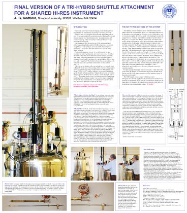
shuttler consists of a linear motor (near left)
based on two pulleys driven by a large stepper
motor two long timing belts driven by the
pulleys and extending up 1 meter over two idler
pulleys and a cross piece coupling the belts to
a push rod extending down into the magnet bore to
which the sample tube is attached (see the bottom
of the poster). For years I felt that any linear
motor, used in this way, would vibrate/oscillate
at the end of its travel which would modulate and
destroy the NMR signal. Finally I invented a
solution to this problem, inspired by the design
of the first, all-pneumatic, version of our
device. There the 5 or 8 mm sample tube is
attached to a freely moving 21 mm diameter
shuttle confined to the inside of a precision
glass tube. The shuttle comes to a stop at a
shoulder below the bottom of the glass tube,
cushioned by a pile of O-rings, so that the
sample is in the exact sensitive region. Now, we
use a pseudo-shuttle that looks similar to the
old one at the bottom, but is constrained at the
top to be able to move 8 mm relative to the push
rod. The photos at the right show the shuttle at
the two extreme positions, and below right is a
cartoon of the coupling assembly, inside the
precision glass tube, seated on the lower support
of the glass shuttle tube. The upper and lower
parts of the assembly are shaded /// and \\\
respectively and actually consist of several
sub-parts screwed or cemented together. We
then program the stepper motor to stop just (3
mm) below the point where the shuttler is
constrained to stop by the shoulder at
the bottom, and the NMR sample is precisely at
the sensitive region of the probe (lower right).
The NMR sample is sucked up to a defined
position during the relax-time by application of
air suction, and is held down securely by air
pressure during readout, isolated from vibration
of the linear motor, by means of the same
pressure/suction system left over from the
previous all-pneumatic system. It works!!
INTRODUCTION For several years I have been
developing a field-shuttling device that rolls
into an instrument room and up to a Varian 500
NMR. Within an hour we can replace the probe
and upper tube with our own Varian-equivalent
probe and precision glass shuttle tube, set up a
frame to support our current linear motor, mount
the motor, and connect things up. After several
days of data production we can disassemble and be
gone. Our attachment could be used in any NMR
instrument (most probably including Bruker and
Jeol NMRs) that is in a room with enough head-
room and that is not restricted by the continuous
presence of a cryoprobe. (this latter restriction
is not ours but is purely paranoia). The first
all-pneumatic version (1) was laborious to use
and denatured proteins. A preliminary motorized
version was described (ICMRBS 2006) that did not
denature proteins but was limited in field range
it still used pneumatics as well as a stepper
motor in conjunction with our key invention of a
special linkage (below), and used only a single
belt. The double belt used here reduces
large-scale vibrations which are a problem, but
not fatal, for H2O solvent (HSQC). Our work on
the shuttler will soon end and here we describe
what will shortly be our final version using a
third level of cycling, namely an electrical
final stage using a small solenoid to demagnetize
from about .035 T to 0.002 T within millisecs,
as previously used long ago by the labs of Hahn
and Pines. Without this new coil our current
attained speed is about 120 msec to about 1 T and
about 250 msec to .003 T. The greater speed at
very low fields is needed for interesting
membrane work on larger vesicles. Our cycler is
is now completely automated. The essential
feature of our final version are described here,
and will be published (in part on the web). This
device is completely unique in its wide field
range, versatility, portability, and
replicability.
What is high resolution shuttling? It is any
shuttling experiment using a high-resolution high
field magnet for detection and preparation. The
first version of our instrument was an
improvement an earlier version from Bryants lab
(2). I got the idea of using a timing-belt-driven
shuttler from the device built in Vieths lab. I
use a commercial system to save time but soon
realized that by doing so I would greatly
enhance the ability of others to duplicate the
device. Unfortunately it has not yet been
duplicated, and no commercial vendor is as yet
interested. Why shuttle? Provided T1 for the
observed nucleus is long enough (0.2 sec or
more) it provides the most sensitive, well
resolved, way to study R1 over the entire range
below 11.7 T, vastly superior to use of lower
field instruments now only available in a few
places, or to electronically switched systems
(commercially available from STELAR, Italy)
except for speed. We have taken (3) interesting
2D R1 data on 15N in a fully labeled protein in
H2O, but this use is limited to above about 4 T
because the geminal proton on the nitrogen
relaxes the nitrogen too rapidly at lower fields
than 4T, for our equipment. The method has
been demonstrated extensively for use by us with
31P in nucleic acids (4) and membranes (5-11)
(see also my other poster with Mingming Pu and
Mary Roberts), and more recently for 13C
carbonyls in membranes (look for another poster
here by Siva Natarajan). These nuclei will be
usable for proteins, to get interesting distances
from them to protons, or to engineered spin
labels or natural paramagnets. A vast future area
of application is likely use small molecules in
fast exchange with binding sites on larger
molecules, with or without spin labels, including
use of small binders as reporters of paramagnetic
relaxation (2) or other properties. Bryant
pointed out the great versatility of cycling for
this purpose since the concentration of the
relaxing species can be varied to match T1 of
the observed species to the speed of the
shuttler. Other possibilities are to characterize
more potentially interesting features now likely
to be hidden in nominally disordered regions of
proteins (3) and to detect weak binding between
small objects (proteins or lipids (11) ) and
larger aggregates, and to detect dynamics in the
now-unobservable 1 microsec to 10 nanosec
time-scale in small proteins (ask me how!).
Shown at the extreme right is the entire device
mounted on the magnet. It takes less than one
hour to remove the normal probe and top tube,
and install our entire system including our own
Varian probes. The photo shows only the edge of
the solenoid valve manifold, which is mounted on
a rolling relay rack, and the rubber tube that
transmits air pressure or suction to the shuttle
tube. This rack (not shown) also holds three
ballast tanks and pressure valves for two levels
of pressure (0.15 bar and 0.3 bar), and 0.2
bar suction. We also do not show another wheeled
rack containing power supplies for the coils, and
a rolling table with an oscilloscope. Raunchy
electronics is distributed on the racks and
table, soon to be replaced with a PC and
microprocessors. In the upper left inset the
linear motor assembly is shown. It is clamped
onto the frame (just below, right) during
running, or else on a Redfield-made rope-operated
fork lift assembly when the apparatus is stored,
visible (below, center) as we roll it up to the
magnet and slide it over to the magnet bore
(below, center left). I built the fork lift to
make it unnecessary to find 2 strong grad
students to lift the 11 Kg motor to the rail, 2
meters above the floor. The motor assembly is
based on a 12.7 mm-thick plate 50 mm high and
220 mm long to which are attached two similarly
heavy perpendicular plates, one of which supports
the stepper motor (the heaviest we thought we
could handle). Two lighter plates, parallel to
the end plates, hold ball bearings for the shaft
from the motor. To this shaft are clamped the two
lower timing belt pulleys that are 45 mm in
diameter. The upper smaller idler pulleys are
each held by an assembly attached to a lever arm
that sits on a grooved fulcrum. These fulcrums
are supported by single a support which is heavy
at the bottom and light at the top. A system of
cables, springs, pulleys, and turnbuckles exerts
force (10 KgF) on the back end of the lever
arms, to provide equal tension on the two
belts. Thanks to Frank Mello, our machinist!
Our Publications Besides (1,3) and our other
two posters here we have (4), preliminary study
of a nucleic acid (DNA octamer) with good
separation of dipolar and two CSA time-scales
for 31P, showing different degrees of picosec
motion for different bases in this monotonous
molecule (5), first extensive report on 31P
dynamics in membrane vesicles and micelles down
to 0.1 T showing, in addition to effects seen in
(4), evidence for internal motion affecting the
head group in a membrane (6), first extension
of observations down to nearly zero T, giving a
well defined dispersion below 0.05 T that is
readily interpretable in terms of an average
Hamiltonian (7), a report of a state-of-the-art
computer simulation, by others, of a
phospholipid membrane, for which we contributed
high temperature data for comparison, with good
agreement (8, 9), studies of effect of
temperature, vesicle composition, cholesterol,
and binding of a peripheral membrane enzyme, on
dynamics and other properties (10), assaying
sizes of large complexes of proteins and
micelles and (11), mainly biochemistry but
includes application of the method to determine
free-lipid for a lipid/micelle equilibrium in a
complex mixture. This poster will be amplified
for publication and a lengthy web posting will
appear with even more details, as I posted for
ref (1) (go to life sciences section
of www.brandeis.edu and look for my web site). I
thank my collaborators (below) for producing
interesting results and writing them up for
publication while I engineered this device.
Thanks to NIH GM077974 for support, and NSF Chem.
Equipment Div. and ACS PRF fund for earlier
crucial support, as well as TIAA CREF.
Shown below are the glass shuttle tube that
replaces the usual upper stack tube provided by
Varian and (below it) the entire push-rod
assembly. The left end of the tube assembly has
spacers that fit the inside of the shim tube and
the probe upper end, and align them. The
transparent lower section is unused, while the
transparent upper section (right of photo) will
support the small high-speed coil centered 63 mm
above the magnet top flange. The sample is sealed
(1) in a short (3.4) 8 mm NMR tube which is in
turn, cemented into a short plastic holder (1)
that screws into the bottom of the
pseudo-shuttle.
Shown left the upper ends of the shuttle tube
and push rod, and the coils. The Helmholz coil
assembly sits directly on the magnet, with its
top just below the level of the frame. Then the
shuttle tube assembly slides into it and is
supported vertically by it. The glass cylinder on
the push rod is a precision extension of the main
shuttle tube, with a spigot on top for air
pressure/vacuum and a low-friction slinky seal to
the rod on top. A small fast coil will live on
the upper end of the glass tube and be powered
(hopefully) by a commercial supply (Kepco BOP
(Diz lives!)).
References 1. AGR, MRC 41753-768 (03). 2. K.
Victor, A.Van-Quyn, and R. Bryant, Biophys. J.,
88443-454 (2005). 3. J. Clarkson, E.
Eisenmesser, D. Kern et Al, unpublished poster at
07 ISMAR, indicating that a disordered loop
may in fact be a more or less organized structure
moving as one piece. 4. M. F. Roberts (MFR) et.
al., JACS (04). 5. MFR AGR, JACS
12513765-13777 (04). 6. MFR and AGR, PNAS 104
17066-17071 (04) . 7. J. Klauda et al., Biophys.
J. 943074-3083 (08). 8. M. Pu, X. Fang, AGR,
A. Gershenson, MFR, (submitted) (comparison of
activity, binding (by light scattering) and NMR
rxn effects vs lipid composition). 9. MFR, U.
Mohanty, AGR, (in prep). (temperature and
cholesterol effects on lipid dynamics). 10. Y.
Shi, C. Shao, C. Zambonelli, AGR, J. Head, B.
Seaton, MFR, JBC (in press) (08). 11. Y Wong et
al., JACS 1307746 (08).


