
The Lymph System - PowerPoint PPT Presentation
1 / 137
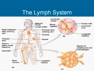
Title: The Lymph System
1
The Lymph System
2
Lymphatic System Overview
- Consists of two semi-independent parts
- A meandering network of lymphatic vessels
- Lymphoid tissues and organs scattered throughout
the body - Returns interstitial fluid and leaked plasma
proteins back to the blood - Lymph interstitial fluid once it has entered
lymphatic vessels
3
(No Transcript)
4
Lymphatic Vessels
- A one-way system in which lymph flows toward the
heart - Lymph vessels include
- Microscopic, permeable, blind-ended capillaries
- Lymphatic collecting vessels
- Trunks and ducts
5
Lymphatic Capillaries
- Similar to blood capillaries, with modifications
- Remarkably permeable
- Loosely joined endothelial minivalves
- Withstand interstitial pressure and remain open
- The minivalves function as one-way gates that
- Allow interstitial fluid to enter lymph
capillaries - Do not allow lymph to escape from the capillaries
6
Lymphatic Capillaries
- During inflammation, lymph capillaries can
absorb - Cell debris
- Pathogens
- Cancer cells
- Cells in the lymph nodes
- Cleanse and examine this debris
- Lacteals specialized lymph capillaries present
in intestinal mucosa - Absorb digested fat and deliver chyle to the
blood
7
Lymphatic Trunks
- Lymphatic trunks are formed by the union of the
largest collecting ducts - Major trunks include
- Paired lumbar, bronchomediastinal, subclavian,
and jugular trunks - A single intestinal trunk
8
Lymphatic Trunks
- Lymph is delivered into one of two large trunks
- Right lymphatic duct drains the right upper arm
and the right side of the head and thorax - Thoracic duct arises from the cisterna chyli
and drains the rest of the body
9
Lymph Transport
- The lymphatic system lacks an organ that acts as
a pump - Vessels are low-pressure conduits
- Uses the same methods as veins to propel lymph
- Pulsations of nearby arteries
- Contractions of smooth muscle in the walls of the
lymphatics
10
Lymph Video
11
Lymphoid Cells
- Lymphocytes are the main cells involved in the
immune response - The two main varieties are T cells and B cells
12
Lymphocytes
- T cells and B cells protect the body against
antigens - Antigen anything the body perceives as foreign
- Bacteria and their toxins viruses
- Mismatched RBCs or cancer cells
13
Lymphocytes
- T cells
- Manage the immune response
- Attack and destroy foreign cells
- B cells
- Produce plasma cells, which secrete antibodies
- Antibodies immobilize antigens
14
Other Lymphoid Cells
- Macrophages phagocytize foreign substances and
help activate T cells - Dendritic cells spiny-looking cells with
functions similar to macrophages - Reticular cells fibroblastlike cells that
produce a stroma, or network, that supports other
cell types in lymphoid organs
15
Lymph Nodes
- Lymph nodes are the principal lymphoid organs of
the body - Nodes are imbedded in connective tissue and
clustered along lymphatic vessels - Aggregations of these nodes occur near the body
surface in inguinal, axillary, and cervical
regions of the body
16
Lymph Nodes
- Their two basic functions are
- Filtration macrophages destroy microorganisms
and debris - Immune system activation monitor for antigens
and mount an attack against them
17
Structure of a Lymph Node
- Nodes are bean shaped and surrounded by a fibrous
capsule - Trabeculae extended inward from the capsule and
divide the node into compartments - Nodes have two histologically distinct regions a
cortex and a medulla
18
Structure of a Lymph Node
- The cortex contains follicles with germinal
centers, heavy with dividing B cells - Dendritic cells nearly encapsulate the follicles
- The deep cortex houses T cells in transit
- T cells circulate continuously among the blood,
lymph nodes, and lymphatic stream
19
Structure of a Lymph Node
- Medullary cords extend from the cortex and
contain B cells, T cells, and plasma cells - Throughout the node are lymph sinuses
crisscrossed by reticular fibers - Macrophages reside on these fibers and
phagocytize foreign matter
20
Structure of a Lymph Node
21
Other Lymphoid Organs
- The spleen, thymus gland, and tonsils
- Peyers patches and bits of lymphatic tissue
scattered in connective tissue - All are composed of reticular connective tissue
and all help protect the body - Only lymph nodes filter lymph
22
Spleen
- Largest lymphoid organ, located on the left side
of the abdominal cavity beneath the diaphragm - It extends to curl around the anterior aspect of
the stomach - It is served by the splenic artery and vein,
which enter and exit at the hilus - Functions
- Site of lymphocyte proliferation
- Immune surveillance and response
- Cleanses the blood
23
Additional Spleen Functions
- Stores breakdown products of RBCs for later reuse
- Spleen macrophages salvage and store iron for
later use by bone marrow - Site of fetal erythrocyte production (normally
ceases after birth) - Stores blood platelets
24
Structure of the Spleen
25
Thymus
- A bilobed organ that secrets hormones (thymosin
and thymopoietin) that cause T lymphocytes to
become immunocompetent - The size of the thymus varies with age
- In infants, it is found in the inferior neck and
extends into the mediastinum where it partially
overlies the heart - It increases in size and is most active during
childhood - It stops growing during adolescence and then
gradually atrophies
26
Thymus
- The thymus differs from other lymphoid organs in
important ways - It functions strictly in T lymphocyte maturation
- It does not directly fight antigens
- The stroma of the thymus consists of star-shaped
epithelial cells (not reticular fibers) - These star-shaped thymocytes secrete the hormones
that stimulate lymphocytes to become
immunocompetent
27
Tonsils
- Simplest lymphoid organs form a ring of
lymphatic tissue around the pharynx - Location of the tonsils
- Palatine tonsils either side of the posterior
end of the oral cavity - Lingual tonsils lie at the base of the tongue
- Pharyngeal tonsil posterior wall of the
nasopharynx - Tubal tonsils surround the openings of the
auditory tubes into the pharynx
28
Aggregates of Lymphoid Follicles
- Peyers patches isolated clusters of lymphoid
tissue, similar to tonsils - Found in the wall of the distal portion of the
small intestine - Similar structures are found in the appendix
- Peyers patches and the appendix
- Destroy bacteria, preventing them from breaching
the intestinal wall - Generate memory lymphocytes for long-term
immunity
29
MALT
- MALT mucosa-associated lymphatic tissue is
composed of - Peyers patches, tonsils, and the appendix
(digestive tract) - Lymphoid nodules in the walls of the bronchi
(respiratory tract) - MALT protects the digestive and respiratory
systems from foreign matter
30
Immunity Two Intrinsic Defense Systems
- Innate (nonspecific) system responds quickly and
consists of - First line of defense intact skin and mucosae
prevent entry of microorganisms - Second line of defense antimicrobial proteins,
phagocytes, and other cells - Inhibit spread of invaders throughout the body
- Inflammation is its hallmark and most important
mechanism
31
(No Transcript)
32
Non-Specific Defense
- What is the first line of defense?
- The skin and mucous membranes
- Protective proteins (lysozyme)
- What is the second line of defense?
- Phagocytic WBCs neutrophylls
- Monocytes macrophages
- Eosinophils fight against parasitic invaders
- Inflammatory response
- Compliment
- interferons
33
Immunity Two Intrinsic Defense Systems
- Adaptive (specific) defense system
- Third line of defense mounts attack against
particular foreign substances - Takes longer to react than the innate system
- Works in conjunction with the innate system
34
Surface Barriers
- Skin, mucous membranes, and their secretions make
up the first line of defense - Keratin in the skin
- Presents a formidable physical barrier to most
microorganisms - Is resistant to weak acids and bases, bacterial
enzymes, and toxins - Mucosae provide similar mechanical barriers
35
Epithelial Chemical Barriers
- Epithelial membranes produce protective chemicals
that destroy microorganisms - Skin acidity (pH of 3 to 5) inhibits bacterial
growth - Sebum contains chemicals toxic to bacteria
- Stomach mucosae secrete concentrated HCl and
protein-digesting enzymes - Saliva and lacrimal fluid contain lysozyme
- Mucus traps microorganisms that enter the
digestive and respiratory systems
36
Respiratory Tract Mucosae
- Mucus-coated hairs in the nose trap inhaled
particles - Mucosa of the upper respiratory tract is ciliated
- Cilia sweep dust- and bacteria-laden mucus away
from lower respiratory passages
37
Internal Defenses Cells and Chemicals
- The body uses nonspecific cellular and chemical
devices to protect itself - Phagocytes and natural killer (NK) cells
- Antimicrobial proteins in blood and tissue fluid
- Inflammatory response enlists macrophages, mast
cells, WBCs, and chemicals - Harmful substances are identified by surface
carbohydrates unique to infectious organisms
38
White Blood Cells Video
39
Phagocytes
- Macrophages are the chief phagocytic cells
- Free macrophages wander throughout a region in
search of cellular debris - Kupffer cells (liver) and microglia (brain) are
fixed macrophages - Neutrophils become phagocytic when encountering
infectious material - Eosinophils are weakly phagocytic against
parasitic worms - Mast cells bind and ingest a wide range of
bacteria
40
Mechanism of Phagocytosis
- Microbes adhere to the phagocyte
- Pseudopods engulf the particle (antigen) into a
phagosome - Phagosomes fuse with a lysosome to form a
phagolysosome - Invaders in the phagolysosome are digested by
proteolytic enzymes - Indigestible and residual material is removed by
exocytosis
41
Mechanism of Phagocytosis
42
Natural Killer (NK) Cells
- Cells that can lyse and kill cancer cells and
virus-infected cells - Natural killer cells
- Are a small, distinct group of large granular
lymphocytes - React nonspecifically and eliminate cancerous and
virus-infected cells - Kill their target cells by releasing perforins
and other cytolytic chemicals - Secrete potent chemicals that enhance the
inflammatory response
43
Inflammation Tissue Response to Injury
- The inflammatory response is triggered whenever
body tissues are injured - Prevents the spread of damaging agents to nearby
tissues - Disposes of cell debris and pathogens
- Sets the stage for repair processes
- The four cardinal signs of acute inflammation are
redness, heat, swelling, and pain
44
Inflammation Response
- Begins with a flood of inflammatory chemicals
released into the extracellular fluid - Inflammatory mediators
- Include kinins, prostaglandins (PGs), complement,
and cytokines - Are released by injured tissue, phagocytes,
lymphocytes, and mast cells - Cause local small blood vessels to dilate,
resulting in hyperemia
45
Toll-like Receptors (TLRs)
- Macrophages and cells lining the gastrointestinal
and respiratory tracts bear TLRs - TLRs recognize specific classes of infecting
microbes - Activated TLRs trigger the release of cytokines
that promote inflammation
46
Inflammatory Response Vascular Permeability
- Chemicals liberated by the inflammatory response
increase the permeability of local capillaries - Exudate (fluid containing proteins, clotting
factors, and antibodies) - Seeps into tissue spaces causing local edema
(swelling), which contributes to the sensation of
pain
47
Inflammatory Response Edema
- The surge of protein-rich fluids into tissue
spaces (edema) - Helps to dilute harmful substances
- Brings in large quantities of oxygen and
nutrients needed for repair - Allows entry of clotting proteins, which prevents
the spread of bacteria
48
Inflammatory Response Phagocytic Mobilization
- Occurs in four main phases
- Leukocytosis neutrophils are released from the
bone marrow in response to leukocytosis-inducing
factors released by injured cells - Margination neutrophils cling to the walls of
capillaries in the injured area - Diapedesis neutrophils squeeze through
capillary walls and begin phagocytosis - Chemotaxis inflammatory chemicals attract
neutrophils to the injury site
49
Inflammatory Response Phagocytic Mobilization
50
Flowchart of Events in Inflammation
51
Interferon (IFN)
- Genes that synthesize IFN are activated when a
host cell is invaded by a virus - Interferon molecules leave the infected cell and
enter neighboring cells - Interferon stimulates the neighboring cells to
activate genes for PKR (an antiviral protein) - PKR nonspecifically blocks viral reproduction in
the neighboring cell
52
Fever
- Abnormally high body temperature in response to
invading microorganisms - The bodys thermostat is reset upwards in
response to pyrogens, chemicals secreted by
leukocytes and macrophages exposed to bacteria
and other foreign substances
53
Fever
- High fevers are dangerous as they can denature
enzymes - Moderate fever can be beneficial, as it causes
- The liver and spleen to sequester iron and zinc
(needed by microorganisms) - An increase in the metabolic rate, which speeds
up tissue repair
54
Specific Immunity
- What cells are involved in the immune defense ?
- B cells
- T cells
- Macrophages
- What 2 types of immune responses can be mounted?
- Humoral immune response (circulates antibodies
in blood) - Cell-mediated immune response
55
Adaptive (Specific) Defenses
- The adaptive immune system is a functional system
that - Recognizes specific foreign substances
- Acts to immobilize, neutralize, or destroy
foreign substances - Amplifies inflammatory response and activates
complement
56
Adaptive Immune Defenses
- The adaptive immune system is antigen-specific,
systemic, and has memory - It has two separate but overlapping arms
- Humoral, or antibody-mediated immunity
- Cellular, or cell-mediated immunity
57
Antigens
- Substances that can mobilize the immune system
and provoke an immune response - The ultimate targets of all immune responses are
mostly large, complex molecules not normally
found in the body (nonself)
58
Cells of the Adaptive Immune System
- Two types of lymphocytes
- B lymphocytes oversee humoral immunity
- T lymphocytes non-antibody-producing cells that
constitute the cell-mediated arm of immunity - Antigen-presenting cells (APCs)
- Do not respond to specific antigens
- Play essential auxiliary roles in immunity
59
Lymphocytes
- Immature lymphocytes released from bone marrow
are essentially identical - Whether a lymphocyte matures into a B cell or a T
cell depends on where in the body it becomes
immunocompetent - B cells mature in the bone marrow
- T cells mature in the thymus
60
T Cell Selection in the Thymus
61
T Cells
- T cells mature in the thymus under negative and
positive selection pressures - Negative selection eliminates T cells that are
strongly anti-self - Positive selection selects T cells with a weak
response to self-antigens, which thus become both
immunocompetent and self-tolerant
62
B Cells
- B cells become immunocompetent and self-tolerant
in bone marrow - Some self-reactive B cells are inactivated
(anergy) while others are killed - Other B cells undergo receptor editing in which
there is a rearrangement of their receptors
63
Antigen-Presenting Cells (APCs)
- Major rolls in immunity are
- To engulf foreign particles
- To present fragments of antigens on their own
surfaces, to be recognized by T cells - Major APCs are dendritic cells (DCs),
macrophages, and activated B cells - The major initiators of adaptive immunity are
DCs, which actively migrate to the lymph nodes
and secondary lymphoid organs and present
antigens to T and B cells
64
Adaptive Immunity Summary
- Two-fisted defensive system that uses
lymphocytes, APCs, and specific molecules to
identify and destroy nonself particles - Its response depends upon the ability of its
cells to - Recognize foreign substances (antigens) by
binding to them - Communicate with one another so that the whole
system mounts a response specific to those
antigens
65
Humoral Immunity Response
- Antigen challenge first encounter between an
antigen and a naive immunocompetent cell - Takes place in the spleen or other lymphoid organ
- If the lymphocyte is a B cell
- The challenging antigen provokes a humoral immune
response - Antibodies are produced against the challenger
66
Clonal Selection
- Stimulated B cell growth forms clones bearing the
same antigen-specific receptors - A naive, immunocompetent B cell is activated when
antigens bind to its surface receptors and
cross-link adjacent receptors - Antigen binding is followed by receptor-mediated
endocytosis of the cross-linked antigen-receptor
complexes - These activating events, plus T cell
interactions, trigger clonal selection
67
Clonal Selection
68
Fate of the Clones
- Most clone cells become antibody-secreting plasma
cells - Plasma cells secrete specific antibody at the
rate of 2000 molecules per second
69
Fate of the Clones
- Secreted antibodies
- Bind to free antigens
- Mark the antigens for destruction by specific or
nonspecific mechanisms - Clones that do not become plasma cells become
memory cells that can mount an immediate response
to subsequent exposures of the same antigen
70
Immunological Memory
- Primary immune response cellular
differentiation and proliferation, which occurs
on the first exposure to a specific antigen - Lag period 3 to 6 days after antigen challenge
- Peak levels of plasma antibody are achieved in 10
days - Antibody levels then decline
71
Immunological Memory
- Secondary immune response re-exposure to the
same antigen - Sensitized memory cells respond within hours
- Antibody levels peak in 2 to 3 days at much
higher levels than in the primary response - Antibodies bind with greater affinity, and their
levels in the blood can remain high for weeks to
months
72
Primary and Secondary Humoral Responses
73
Active Humoral Immunity
- B cells encounter antigens and produce antibodies
against them - Naturally acquired response to a bacterial or
viral infection - Artificially acquired response to a vaccine of
dead or attenuated pathogens - Vaccines spare us the symptoms of disease, and
their weakened antigens provide antigenic
determinants that are immunogenic and reactive
74
Passive Humoral Immunity
- Differs from active immunity in the antibody
source and the degree of protection - B cells are not challenged by antigens
- Immunological memory does not occur
- Protection ends when antigens naturally degrade
in the body - Naturally acquired from the mother to her fetus
via the placenta - Artificially acquired from the injection of
serum, such as gamma globulin
75
Types of Acquired Immunity
76
MHCs
- What are MHCs?
- Surface glycoproteins that establish self
- Class I MHC found on all nucleated cells
- Class II MHC found on macrophages, B cells and
activated T cells
77
Clonal Selection of B cells
- What is clonal selection?
78
Memory
- How does a secondary immune response differ from
the primary response?
79
Role of Helper T Cells
80
Cell mediated Immunity
81
Humoral Immunity
82
Antibody Actions
83
(No Transcript)
84
Antibody Structure and Function
- Antibodies have variable binding sites
- They are antigen specific
- Monoclonal antibodies use antibody specificity
for diagnosis, and treatment of diseases
85
Antibodies
- Also called immunoglobulins
- Constitute the gamma globulin portion of blood
proteins - Are soluble proteins secreted by activated B
cells and plasma cells in response to an antigen - Are capable of binding specifically with that
antigen - There are five classes of antibodies IgD, IgM,
IgG, IgA, and IgE
86
Classes of Antibodies
- IgD monomer attached to the surface of B cells,
important in B cell activation - IgM pentamer released by plasma cells during
the primary immune response - IgG monomer that is the most abundant and
diverse antibody in primary and secondary
response crosses the placenta and confers
passive immunity - IgA dimer that helps prevent attachment of
pathogens to epithelial cell surfaces - IgE monomer that binds to mast cells and
basophils, causing histamine release when
activated
87
Basic Antibody Structure
- Consists of four looping polypeptide chains
linked together with disulfide bonds - Two identical heavy (H) chains and two identical
light (L) chains - The four chains bound together form an antibody
monomer - Each chain has a variable (V) region at one end
and a constant (C) region at the other - Variable regions of the heavy and light chains
combine to form the antigen-binding site
88
Basic Antibody Structure
89
Antibody Structure
- Antibodies responding to different antigens have
different V regions but the C region is the same
for all antibodies in a given class - C regions form the stem of the Y-shaped antibody
and - Determine the class of the antibody
- Serve common functions in all antibodies
- Dictate the cells and chemicals that the antibody
can bind to - Determine how the antibody class will function in
elimination of antigens
90
Mechanisms of Antibody Diversity
- Plasma cells make over a billion different types
of antibodies - Each cell, however, only contains 100,000 genes
that code for these polypeptides - To code for this many antibodies, somatic
recombination takes place - Gene segments are shuffled and combined in
different ways by each B cell as it becomes
immunocompetent - Information of the newly assembled genes is
expressed as B cell receptors and as antibodies
91
Antibody Diversity
- Random mixing of gene segments makes unique
antibody genes that - Code for H and L chains
- Account for part of the variability in antibodies
- V gene segments, called hypervariable regions,
mutate and increase antibody variation - Plasma cells can switch H chains, making two or
more classes with the same V region
92
Antibody Targets
- Antibodies themselves do not destroy antigen
they inactivate and tag it for destruction - All antibodies form an antigen-antibody (immune)
complex - Defensive mechanisms used by antibodies are
neutralization, agglutination, precipitation, and
complement fixation
93
Complement Fixation and Activation
- Complement fixation is the main mechanism used
against cellular antigens - Antibodies bound to cells change shape and expose
complement binding sites - This triggers complement fixation and cell lysis
- Complement activation
- Enhances the inflammatory response
- Uses a positive feedback cycle to promote
phagocytosis - Enlists more and more defensive elements
94
Other Mechanisms of Antibody Action
- Neutralization antibodies bind to and block
specific sites on viruses or exotoxins, thus
preventing these antigens from binding to
receptors on tissue cells
95
Other Mechanisms of Antibody Action
- Agglutination antibodies bind the same
determinant on more than one antigen - Makes antigen-antibody complexes that are
cross-linked into large lattices - Cell-bound antigens are cross-linked, causing
clumping (agglutination) - Precipitation soluble molecules are
cross-linked into large insoluble complexes
96
Mechanisms of Antibody Action
97
Monoclonal Antibodies
- Commercially prepared antibodies are used
- To provide passive immunity
- In research, clinical testing, and treatment of
certain cancers - Monoclonal antibodies are pure antibody
preparations - Specific for a single antigenic determinant
- Produced from descendents of a single cell
98
Monoclonal Antibodies
- Hybridomas cell hybrids made from a fusion of a
tumor cell and a B cell - Have desirable properties of both parent cells
indefinite proliferation as well as the ability
to produce a single type of antibody
99
Cell-Mediated Immune Response
- Since antibodies are useless against
intracellular antigens, cell-mediated immunity is
needed - Two major populations of T cells mediate cellular
immunity - CD4 cells (T4 cells) are primarily helper T cells
(TH) - CD8 cells (T8 cells) are cytotoxic T cells (TC)
that destroy cells harboring foreign antigens - Other types of T cells are
- Suppressor T cells (TS)
- Memory T cells
100
Importance of Cellular Response
- T cells recognize and respond only to processed
fragments of antigen displayed on the surface of
body cells - T cells are best suited for cell-to-cell
interactions, and target - Cells infected with viruses, bacteria, or
intracellular parasites - Abnormal or cancerous cells
- Cells of infused or transplanted foreign tissue
101
Cytokines
- Mediators involved in cellular immunity,
including hormonelike glycoproteins released by
activated T cells and macrophages - Some are co-stimulators of T cells and T cell
proliferation - Interleukin 1 (IL-1) released by macrophages
co-stimulates bound T cells to - Release interleukin 2 (IL-2)
- Synthesize more IL-2 receptors
102
Cytokines
- IL-2 is a key growth factor, which sets up a
positive feedback cycle that encourages activated
T cells to divide - It is used therapeutically to enhance the bodys
defenses against cancer - Other cytokines amplify and regulate immune and
nonspecific responses
103
Cytokines
- Examples include
- Perforin and lymphotoxin cell toxins
- Gamma interferon enhances the killing power of
macrophages - Inflammatory factors
104
Helper T Cells (TH)
- Regulatory cells that play a central role in the
immune response - Once primed by APC presentation of antigen, they
- Chemically or directly stimulate proliferation of
other T cells - Stimulate B cells that have already become bound
to antigen - Without TH, there is no immune response
105
Helper T Cell
- TH cells interact directly with B cells that have
antigen fragments on their surfaces bound to MHC
II receptors - TH cells stimulate B cells to divide more rapidly
and begin antibody formation - B cells may be activated without TH cells by
binding to T cellindependent antigens - Most antigens, however, require TH co-stimulation
to activate B cells - Cytokines released by TH amplify nonspecific
defenses
106
Cytotoxic T Cell (Tc)
- TC cells, or killer T cells, are the only T cells
that can directly attack and kill other cells - They circulate throughout the body in search of
body cells that display the antigen to which they
have been sensitized - Their targets include
- Virus-infected cells
- Cells with intracellular bacteria or parasites
- Cancer cells
- Foreign cells from blood transfusions or
transplants
107
Cytotoxic T Cells
- Bind to self-antiself complexes on all body cells
- Infected or abnormal cells can be destroyed as
long as appropriate antigen and co-stimulatory
stimuli (e.g., IL-2) are present - Natural killer cells activate their killing
machinery when they bind to MICA receptor - MICA receptor MHC-related cell surface protein
in cancer cells, virus-infected cells, and cells
of transplanted organs
108
Mechanisms of Tc Action
- In some cases, TC cells
- Bind to the target cell and release perforin into
its membrane - In the presence of Ca2 perforin causes cell
lysis by creating transmembrane pores - Other TC cells induce cell death by
- Secreting lymphotoxin, which fragments the target
cells DNA - Secreting gamma interferon, which stimulates
phagocytosis by macrophages
109
Mechanisms of Tc Action
110
Other T Cells
- Suppressor T cells (TS) regulatory cells that
release cytokines, which suppress the activity of
both T cells and B cells - Gamma delta T cells (Tgd) 10 of all T cells
found in the intestines that are triggered by
binding to MICA receptors
111
Summary of the Primary Immune Response
112
Organ Transplants
- The four major types of grafts are
- Autografts graft transplanted from one site on
the body to another in the same person - Isografts grafts between identical twins
- Allografts transplants between individuals that
are not identical twins, but belong to same
species - Xenografts grafts taken from another animal
species
113
Prevention of Rejection
- Prevention of tissue rejection is accomplished by
using immunosuppressive drugs - However, these drugs depress patients immune
system so it cannot fight off foreign agents
114
Immunodeficiencies
- Congenital and acquired conditions in which the
function or production of immune cells,
phagocytes, or complement is abnormal - SCID severe combined immunodeficiency (SCID)
syndromes genetic defects that produce - A marked deficit in B and T cells
- Abnormalities in interleukin receptors
- Defective adenosine deaminase (ADA) enzyme
- Metabolites lethal to T cells accumulate
- SCID is fatal if untreated treatment is with
bone marrow transplants
115
Acquired Immunodeficiencies
- Hodgkins disease cancer of the lymph nodes
leads to immunodeficiency by depressing lymph
node cells - Acquired immune deficiency syndrome (AIDS)
cripples the immune system by interfering with
the activity of helper T (CD4) cells - Characterized by severe weight loss, night
sweats, and swollen lymph nodes - Opportunistic infections occur, including
pneumocystis pneumonia and Kaposis sarcoma
116
AIDS
- Caused by human immunodeficiency virus (HIV)
transmitted via body fluids blood, semen, and
vaginal secretions - HIV enters the body via
- Blood transfusions
- Contaminated needles
- Intimate sexual contact, including oral sex
- HIV
- Destroys TH cells
- Depresses cell-mediated immunity
117
AIDS
- HIV multiplies in lymph nodes throughout the
asymptomatic period - Symptoms appear in a few months to 10 years
- Attachment
- HIVs coat protein (gp120) attaches to the CD4
receptor - A nearby protein (gp41) fuses the virus to the
target cell
118
AIDS
- HIV enters the cell and uses reverse
transcriptase to produce DNA from viral RNA - This DNA (provirus) directs the host cell to make
viral RNA (and proteins), enabling the virus to
reproduce and infect other cells
119
AIDS
- HIV reverse transcriptase is not accurate and
produces frequent transcription errors - This high mutation rate causes resistance to
drugs - Treatments include
- Reverse transcriptase inhibitors (AZT)
- Protease inhibitors (saquinavir and ritonavir)
- New drugs currently being developed that block
HIVs entry to helper T cells
120
HIV Video
121
Autoimmune Diseases
- Loss of the immune systems ability to
distinguish self from nonself - The body produces autoantibodies and sensitized
TC cells that destroy its own tissues - Examples include multiple sclerosis, myasthenia
gravis, Graves disease, Type I (juvenile)
diabetes mellitus, systemic lupus erythematosus
(SLE), glomerulonephritis, and rheumatoid
arthritis
122
Mechanisms of Autoimmune Diseases
- Ineffective lymphocyte programming
self-reactive T and B cells that should have been
eliminated in the thymus and bone marrow escape
into the circulation - New self-antigens appear, generated by
- Gene mutations that cause new proteins to appear
- Changes in self-antigens by hapten attachment or
as a result of infectious damage
123
Mechanisms of Autoimmune Diseases
- If the determinants on foreign antigens resemble
self-antigens - Antibodies made against foreign antigens
cross-react with self-antigens
124
Hypersensitivity
- Immune responses that cause tissue damage
- Different types of hypersensitivity reactions are
distinguished by - Their time course
- Whether antibodies or T cells are the principle
immune elements involved - Antibody-mediated allergies are immediate and
subacute hypersensitivities - The most important cell-mediated allergic
condition is delayed hypersensitivity
125
Immediate Hypersensitivity
- Acute (type I) hypersensitivities begin in
seconds after contact with allergen - Anaphylaxis initial allergen contact is
asymptomatic but sensitizes the person - Subsequent exposures to allergen cause
- Release of histamine and inflammatory chemicals
- Systemic or local responses
126
Immediate Hypersensitivity
- The mechanism involves IL-4 secreted by T cells
- IL-4 stimulates B cells to produce IgE
- IgE binds to mast cells and basophils causing
them to degranulate, resulting in a flood of
histamine release and inducing the inflammatory
response
127
Acute Allergic Response
128
Anaphylaxis
- Reactions include runny nose, itching reddened
skin, and watery eyes - If allergen is inhaled, asthmatic symptoms appear
constriction of bronchioles and restricted
airflow - If allergen is ingested, cramping, vomiting, or
diarrhea occur - Antihistamines counteract these effects
129
Anaphylactic Shock
- Response to allergen that directly enters the
blood (e.g., insect bite, injection) - Basophils and mast cells are enlisted throughout
the body - Systemic histamine releases may result in
- Constriction of bronchioles
- Sudden vasodilation and fluid loss from the
bloodstream - Hypotensive shock and death
- Treatment epinephrine is the drug of choice
130
Immunity in Health and Disease
- How does a vaccination work?
- How is the Rh factor regulated in a pregnant
woman? - What abnormal immune disorders exist?
- Allergic reaction
- Autoimmune disease
- HIV
131
(No Transcript)
132
(No Transcript)
133
(No Transcript)
134
(No Transcript)
135
(No Transcript)
136
(No Transcript)
137
(No Transcript)
Recommended
CrystalGraphics Presentations


