
Acute Kidney Injury (ARF) - PowerPoint PPT Presentation
1 / 46
Title:
Acute Kidney Injury (ARF)
Description:
Acute Kidney Injury (ARF) By: Dr. Hatim Ahmed Hassan Senior Registrar PICU Imaging Ultrasound Useful in Post renal AKI. Early obstruction may not show significant ... – PowerPoint PPT presentation
Number of Views:711
Avg rating:3.0/5.0
Title: Acute Kidney Injury (ARF)
1
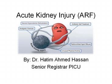
Acute Kidney Injury (ARF)
- By Dr. Hatim Ahmed Hassan
- Senior Registrar PICU
2
Objective
- Introduction and background
- Definition
- Epidemiology
- Physiology
- Etiology
- Clinical presentation
- Diagnosis
- management
3
INTRODUCTION
- AKI is defined as the abrupt loss of kidney
function that results in a decline in GFR,
retention of urea and other nitrogenous waste
products, and dysregulation of extracellular
volume and electrolytes. - The term AKI has largely replaced (ARF), as it
more clearly defines renal dysfunction as a
continuum rather than a discrete finding of
failed kidney function. - Pediatric AKI presents with a wide range of
clinical manifestations from a minimal elevation
in serum creatinine to anuric renal failure,
arises from multiple causes, and occurs in a
variety of clinical settings .
4
Background
- Acute kidney injury (previously known as acute
renal failure) covers a wide spectrum of injury
to the kidneys, not just kidney failure - Up to 18 of all hospital admissions have AKI
- Inpatient AKI-related mortality is between 25 and
30 - Between 20 and 30 of cases of AKI are
preventable. Prevention could save up to 12,000
lives each year - NHS costs related to AKI are between 434 and
620 million per year
5
Definition
- AKI is defined as a decrease in glomerular
filtration rate (GFR), which traditionally is
manifested by an elevated or a rise in serum
creatinine. - However, serum creatinine is often a delayed and
imprecise test as it reflects GFR in individuals
at steady state with stable kidney function, and
does not accurately estimate the GFR in a patient
whose renal function is changing. For example, a
child in the early stages of severe AKI with a
markedly reduced GFR may have a relatively normal
or slightly elevated creatinine, as there has not
been sufficient time for creatinine accumulation..
6
Definition
- In addition, creatinine is removed by dialysis,
and it is not possible to assess renal function
using serum creatinine once dialysis is
initiated. - Despite these limitations, elevated or a rise in
serum creatinine continues to be the most widely
used laboratory finding to make the diagnosis of
AKI in children
7
Definition
8
Definition
9
EPIDEMIOLOGY
- The precise incidence and prevalence of pediatric
acute kidney injury (AKI) are not known, largely
due to the lack of a consensus definition in
published studies. The incidence varies based on
the definition used and potentially geographic
location
10
Epidemiology of AKI
- Community acquired AKI seen in 1 of all
hospitalized patients on admission.50 of those
patients have underlying CKD. - Development of AKI in hospitalized patients is
common and carries independent mortality risk. - In patients with normal renal function, the
incidence of AKI is about 5. - In patients with underlying CKD, the incidence is
about 16
11
Epidemiology of AKI
- Hospital acquired AKI
- 40 is due to ATN
- 15 related to medication associated AKI.
- 10 due to contrast induced nephropathy.
- AIDS associated AKI account for 5.
12
PHYSIOLOGY
13
Types of AKI
- AKI
- AKI/CKD
- Anuric (lt50ml of urine output/day)
- Oliguric (lt400 ml/day)
- Non-oliguric (gt400 ml/day)
14
ARF Pirouz Daeihagh, M.D.Internal
medicine/Nephrology Wake Forest University
School of Medicine. Downloaded 4.6.09
15
Etiology of AKI
- Prerenal
- Renal hypoperfusion, no structural damage to
the kidneys, Cr normalizes in 24-72 hours with
correction of hypoperfused state. - Post-renal
- Obstruction to the urine flow, either
unilateral/bilateral, intra-ureteral or
extra-ureteral or bladder neck or intra-pelvis
(renal pelvis). - Intra-renal
- Damage or inflammation within the kidney, may
be primary renal or part of systemic disease.
16
Prerenal AKI
17
Prerenal AKI
18
Intrarenal hemodynamic changes
19
Intrarenal AKI
- Vascular
- Glomerular
- Interstitial
- Tubular
20
Vascular causes of Intrarenal AKI
- Large and Medium size vessels
- Renal artery thrombosis or emboli
- Renal vein thrombosis
- Polyarterial nodosa
- Small vessel disease
- Atheroembolic phenomenon
- Microangiopathies like TTP, HUS, HELLP and
malignant HTN.
21
Glomerular causes of Intrarenal AKI
- Nephritis
- Hematuria
- Proteinuria (1-2gm/d)
- ARF
- May present as Rapidly progressive
Glomerulonephritis - Renal Biopsy to diagnose
- Nephrosis
- Minimal hematuria
- Massive proteinuria(gt3gm/d)
- Uncommon to present as ARF
- Renal Biopsy not needed to diagnose.
22
Interstitial causes of Intrarenal AKI
- Focal/diffuse edema and infiltration of the renal
interstitium with inflammatory cells.
23
Tubular causes of Intrarenal AKI, Acute Tubular
Necrosis
- Ischemia induced
- Shock
- Hemorrhage
- Sepsis
- Trauma
- Pancreatitis
- Nephrotoxin induced
- Drugs like IV contrast, Aminoglycosides, Ampho B,
pentamidine, Acyclovir, Ehtylene Glycol etc., - Endogenous Toxins in the case of Rhabdomyolysis,
Hemolysis, uric acid nephropathy
24
Postrenal AKI
- Intra Ureteral
- Stones, Clots, Pyogenic debris, Sloughed
papillae in analgesic nephropathy, sickle cell
disease etc., - Extra Ureteral
- Malignancy, Retroperitoneal fibrosis,
accidental ligation etc., - Bladder neck/Urethral
- Autonomic neuropathy with urinary retention,
Urethral stricture, Blood clots/bladder stones.
25
CLINICAL PRESENTATION
- (Symptoms of acute renal failure depend largely
on the underlying cause.) - Fever
- Rash
- Bloody diarrhea
- Severe vomiting
- Abdominal pain
- Hemorrhage
- No urine output or high urine output
- History of recent infection
- Pale skin
26
CLINICAL PRESENTATION
- History of taking certain medications
- History of trauma
- Swelling of the tissues
- Inflammation of the eye
- Detectable abdominal mass
- Exposure to heavy metals or toxic solvents
27
Evaluation of ARF
- Careful History and tabulation of data including
u.o, weights, vitals, medications etc.,. - Physical Examination findings including signs of
vol. depletion etc., - Urinalysis
- Urinary indices(Urine sodium, creatinine, FeNa,
FeUrea etc.,)
28
Mortality associated with AKI
- ICU associated AKI along with respiratory failure
requiring hemodialysis, the mortality is gt90. - ICU associated AKI with out respiratory failure
or hemodialysis, it is 72 - Non-ICU renal failure associated mortality is
around 32.
29
Urinary Indices
- Prerenal
- High SpGr
- No proteinuria/hematuria
- U.Na lt20
- U.Cr/P.Cr gt40
- U.Osm gt500
- FeNa lt1
- FeUrea lt35
- ATN
- Sp Gr 1.010
- Variable proteinuria
- U.Na gt40
- U.Cr/P.Cr lt20
- U.Osm lt350
- FeNa gt1
- FeUrea gt50
30
Urinalysis and Urine Sediment
- UA positive for heme and proteinuria seen in
Glomerular and Interstitial renal failure. - Urine eosinophils are seen in AIN, Atheroembolic
disease etc., - Urine sediment positive for red cell casts seen
in Glomerulonephritis. - UA bland in Post Renal ARF.
31
(No Transcript)
32
Laboratory Data
- .Hypocomplementemia seen in SLE, MPGN,
Atheroembolic disease etc., - Elevated ESR seen in Atheroembolic disease.
- Serologies positive in glomerular diseases, like
ANA, ANCA, Anti GBM, Hepatitis, HIV - Elevated LDH seen in RVT.
33
Laboratory Data (contd)
- Thrombocytopenia with microangiopathic hemolysis
seen in TTP, HUS etc., - Low Haptoglobin, High retic count seen in
microangiopathic states. - Schistocytes (red cell fragmentation).
- CPK, uric acid levels etc., to evaluate for
rhabdomyolysis, uric acid nephropathy. - Evidence of hepatic insufficiency in diagnosing
hepatorenal syndrome.
34
Imaging
- Ultrasound
- Useful in Post renal AKI.
- Early obstruction may not show significant
hydronephrosis. - External obstruction encrasing the whole urinary
system may not show hydronephrosis, for e.g.,
retroperitoneal fibrosis. - U/S doppler useful in diagnosing Renal vein
thrombosis.
35
Imaging (contd)
- CT scan
- Useful for detecting stones, location of the
obstruction, Tumours etc., - Isotope renography
- To evaluate the function significance of
obstruction. - Done with lasix and Mag3 isotope for evaluatine
obstruction.
36
Imaging (contd)
- Cystoscopy and Retrograde Pyelography
- To evaluate patients with high clinical suspicion
of obstruction esp., in unique cases of calculi,
pyogenic debris, blood clots, bladder cancer
etc., - Renal Angigraphy
- In emergent cases of anuria with suspicion of
renal embolization.
37
Renal Biopsy
- Only in patients with no clear etiology.
- In patients with active urinary sediment (RBCs,
red cell casts etc., ) - RPGN (rapidly progressive glomerulonephritis).
- Refractory ATN with out recovery despite no
further renal insults. - Acute Interstitial nephritis.
38
Management of AKI
- Volume repletion with isotonic fluids to improve
renal perfusion pressures in prerenal states. - CVP/ PEWS monitoring.
- Supportive measures for sepsis with pressors,
antibiotics etc., - Colloidal substances like blood products in
hemorrhagic shock. - Management of heart failure by improving cardiac
output.
39
Children and young people ongoing hospital
assessment
- Consider a paediatric early warning score (PEWS)
to identify children and young people at risk of
acute kidney injury - Record physiological observations at admission
and then according to local protocols for given
PEWS - Increase the frequency of observations if
abnormal physiology is detected - Use PEWS with multiple-parameter or aggregate
weighted scoring systems that allow a graded
response and include - heart rate
- respiratory rate
- systolic blood pressure
- level of consciousness
- oxygen saturation
- temperature
- capillary refill time
40
Management (contd)
- Drugs need to be dosed according to the renal
clearance. - Electrolyte and acid base correction.
- Renal diet, if K high.
- Diuretics in overt fluid overload states.
- Foley catheterization in bladder neck
obstruction/prostatic obstruction.
41
(No Transcript)
42
(No Transcript)
43
Management (contd)
- Avoid nephrotoxic agents like Contrast dye,
NSAIDs, Aminoglycosides etc., - Also avoid ACEI unless the underlying problem is
decompensated heart failure. - Nutritional support with parenteral or enteral
feeding.
44
Management (contd)
- Renal replacement therapy
- Modes of dialysis
- IHD (Intermittent Hemodialysis)
- Quick removal of solutes over 3-4 hours,
possible hemodynamic instability. ICU,
hypotensive patients are probably not the best
candiadtes for this type of HD. - CRRT (Continuous renal replacement therapy).
- Modality of choice in critically ill patients.
45
Management (contd)
- Vascular access needed for Hemodialysis.
- Peritoneal dialysis uncommonly used for managing
ARF - It may be used in locations where IHD or CRRT are
not available.
46
Any Questions?